

Chris:
In the 15 y/o patient which was presented with asymmetrical feet, it
appeared
as if the right foot had a significant pes plano-valgus deformity.
The
lengthening osteotomy of the calcaneus which was performed to correct
for the
pes plano-valgus deformity appears to have partially corrected
the clinical
appearance of the right foot. From the photos and EMED analyses,
it appears
as if foot shape and function is improved.
The postoperative radiographs do not appear to be weightbearing.
Therefore,
the postoperative radiographs could not be used to effectively compare
with
the preoperative radiographs which were weightbearing.
In addition, in the
preoperative radiographs, I do not see that much of a hallux abductus
angle
which is normally 10-15 degrees. In the preoperative dorso-plantar
radiographs, I measured about 18 degrees hallux abductus angle on the
left and
about 8 degrees hallux abductus angle on the right. Possibly
Dr. MacWilliams
is using a different measurement protocol for "hallux angle" than what
podiatrists standardly call the "hallux abductus angle" which is
the angle
formed between the bisection of the first metatarsal and the bisection
of the
hallux proximal phalanx.
The questions are:
1. What do you think of the result?
The result appears favorable from a EMED and photo assessment.
However,
clinical result would also be determined by whether the patient
was less
symptomatic and had increased ability to perform weightbearing activities
with
decreased symptoms. Again, radiographs are hard to compare preoperatively
versus postoperatively since the preops were done weightbearing and
the
postops were done nonweightbearing.
2. How do you explain the improvement in arch index & hallux angle?
Again, I don't see that the hallux abductus angle was abnormal to begin
with
on the right foot. Possibly the "hallux angle" is a different
measurement
technique which I am not aware of.
The improvement in arch index is explained by the mechanical effects
of a
calcaneal lengthening osteotomy on the static structure of the foot
in relaxed
bipedal stance. In the right foot preoperatively, there is obvious
gross
medial deviation of the subtalar joint (STJ) axis in relation
to the plantar
structures of the foot. This is apparent especially in the photos
from the
posterior aspect of the foot where there is convexity inferior to the
medial
malleous and from the photo from the anterior aspect of the foot where
there
is convexity along the medial border of the foot. If there is
gross convexity
both inferior to the medial malleolus and along the medial border of
the foot
then this indicates that the talus head has internally rotated and
plantarflexed in relation to the calcaneus. The result
of this abnormal
movement and position of the talus in relation to the calcaneus is
that the
STJ axis becomes internally rotated (i.e. medially deviated)
in relation to
the calcaneus and other plantar structures of the foot.
Since now the weighbearing surface of the foot is mostly lateral to
the STJ
axis due the gross medial deviation of the STJ axis, the ground
reaction force
(GRF) acting on the plantar foot is predominantly lateral to the STJ
axis.
This situation creates an overabundance of pronation moment acting
across the
STJ axis in weightbearing activites which drives the STJ into its
maximally
pronated position. If the STJ has a larger than normal
range of pronation
motion, the result is that seen in the preoperative EMED of the right
foot
where there is an increased shift in plantar pressure toward the
first
metatarsal head and away from the fourth and fifth metatarsal
heads.
The Evans type calcaneal lengthening osteotomy has the effect
to lengthen the
lateral column of the forefoot (including cuboid, 4th and 5th metatarsals)
relative to the medial column of the forefoot (including navicular,
cuneiforms
and 1st, 2nd and 3rd metatarsals). This "pushes" the forefoot
into a more
adducted position in relation to the rearfoot and "pushes"
the rearfoot into a
more medial position in relation to the forefoot. The
result is that the GRF
now acting on the medial calcaneal tubercle of the calcaneus
and on the
metatarsal heads is more medial in relation to the STJ axis than that
which
was present preoperatively. This shifts the GRF from a more lateral
position
in relation to the STJ axis which was present preoperatively to a more
medial
position in relation to the STJ axis which is now present postoperatively.
This relative shift in the position of the weightbearing structures
of the
plantar foot to a more medial position in relation to the STJ axis
creates
increased supination moment and decreased pronation moment
acting across the
STJ axis during weightbearing activities. The biomechanical effect
of surgery
is evidenced in the postoperative EMED with the increased supinated
position
of the STJ being evidenced by the increased weightbearing by the more
lateral
metatarsal heads.
Therefore, the improvement in arch index is explained by the structural
and
functional effects of the calcaneal lengthening osteotomy on the foot.
The
overall effect of the surgery in a flatfoot patient such as described
above is
to externally rotate and laterally translate the STJ axis in
relation to the
plantar weightbearing structures of the foot which causes GRF acting
on the
plantar foot to cause increased supination moment across the STJ axis
during
weightbearing activities.
This is the best I can do with words to explain this concept.
It is much
easier to explain with diagrams. If anyone needs references for
the concepts
outlined above I would be happy to oblige.
Thanks, Chris and Bruce, for the interesting case.
Sincerely,
Kevin
Kevin A. Kirby, D.P.M.
Assistant Clinical Professor of Biomechanics
California College of Podiatric Medicine <Kevkirby@aol.com>
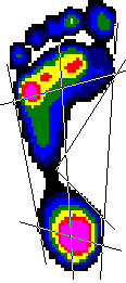
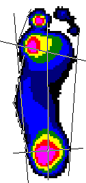
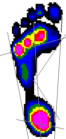
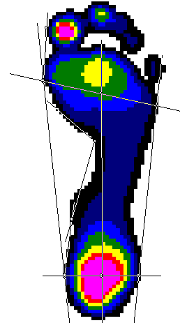
We use a canned package from Novel to compute arch index
and hallux
angle directly from the pressure maps. Lines are fit
to the pressures, and
it is not an exact science. In fact it often gives totally erroneus
predictions, as was the case with the pre-op right foot. In this cases
we
are able to drag nodes to move the lines to where we "think" they should
be
drawn. Arch index is angle between tangents to the medial forefoot
and
hindfoot to the center of the arch. Hallux angle is the tangent
from the
medial aspect of the great toe to the medial aspect of the forefoot,
the
angle that this line makes with the foot axis (mid line of forefoot
and
heel). I've included pre and post-op figures showing these geometric
lines
(as well as others).
Thanks,
Bruce MacWilliams, Ph.D <b.a.macwilliams@m.cc.utah.edu>


Again, the effect of the lengthening osteotomy of the calcaneus is to
lengthen
the lateral column of the foot in relation to the medial column.
The result
is a three dimensional change in foot shape in response to, essentially,
a
transverse plane corrective rearfoot osteotomy.
As an aside, I feel that one of the more important concepts that is
not
discussed frequently enough in regard to plantar pressure measurement
systems
is that the position of the subtalar joint (STJ) axis in relation
to the
plantar foot needs to be considered in order to make assumptions
of how those
ground reaction forces will alter the magnitude of supination and pronation
moments acting across the STJ axis during gait. The child with
the
asymmetrical flatfoot deformity presented by Bruce in this case
is an
excellent example of where it would be very valuable to have the approximate
location of the STJ axis superimposed over the pedobarograph image
to give us
a much better idea of how these plantar pressure images are affecting
the
rotational equilibrium across the STJ axis in static stance, and affecting
the
dynamic moments acting across the STJ axis during gait. I strongly
believe
that combining the STJ axis location along with plantar pressure data
will be
the next step that researchers will need to make to increase our knowledge
in
regard the static and dynamic function of the foot and lower extremity.
Thanks again to Bruce for these wonderful clinical examples and pedobarograph
images.
Sincerely,
Kevin
Kevin A. Kirby, D.P.M. <Kevkirby@aol.com>
Assistant Clinical Professor of Biomechanics
California College of Podiatric Medicine
Private Practice:
2626 N Street
Sacramento, CA 95816
Voice: (916) 456-4768 Fax: (916) 451-6014