|
|
Torsions of the osseous structure of the lower
extremity affect not only the angle of gait but also the function of the
foot. The principle torsions of clinical interest are femoral torsion and
malleolar torsion. The clinical picture of femoral torsion is complicated
by positional (soft tissue) relationships of the hip. This treatise will
describe the normal torsional relationship of the hip first, followed by
the positional relationships of the femur to the pelvis, then the abnormal
torsional relationships of the femur, and finally the torsion of the tibia.
The effects on foot function and gait will be described at the end of the
treatise.
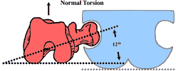
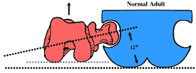
NORMAL TORSION OF THE FEMUR: In the normal adult, the head and neck of the femur are angulated by 12 degrees relative to the femoral condyles as noted in the illustration below.
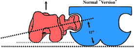
POSITIONAL RELATIONSHIP OF HIP ("VERSION" ANGLES) In the normal adult the Head and Neck of the Femur is angulated approximately 12 degrees relative to the frontal plane of the body. In other words, the angle of the head and neck of the femur is measure relative to the posterior pelvis. Note that the leg faces straight forward. The gluteal fold is drawn for purposes of orientation only and is obviously not anatomically accurate. This represents a normal femur correctly "positioned" into the acetabulum. Note that the angle that the head and neck of the femur makes with the femoral condyles is identical to the angle that the head and neck make with the frontal plane of the body.
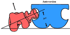
ANTEVERSION is an increase in the angle of the head and neck of the femur relative to the frontal plane of the body. This represents a normal femur abnormally positioned in the acetabulum. The net effect of this positional relationship is an externally rotated leg.
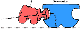
RETROVERSION is a decrease in the angle of the head and neck of the femur relative to the frontal plane of the body. This represents a normal femur that is abnormally positioned relative to the acetabulum. The net effect of this positional relationship is an internally rotated leg.
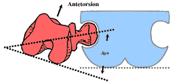
In the three preceeding illustrations the angular relationship between the head and neck of the femur and the femoral condyles (torsion) is normal. These three illustrations describe the positions of a normal femur relative to the frontal plane of the pelvis. In the following description of femoral torsional abnormalities, it is important to understand that the angle that the head and neck of the femur make with the frontal plane of the body is 12 degrees. In other words the position of the head and neck relative to the pelvis is normal but the torsion within the bone is abnormal. ANTETORSION is an increase in the angle of the head and neck of the femur relative to the femoral condyles as noted below. An angle of 30 degrees is shown. Note that the angle that the head and neck of the femur make with the frontal plane of the body is normal and therefore Antetorsion results in an internally deviated thigh.
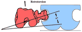
RETROTORSION is a decrease in the angle of the head and neck of the femur relative to the femoral condyles. In the illustration the angle is approximately 8 degrees. Note that the angle that the head and neck of the femur make with the frontal plane of the body is normal and therefore Retrotorsion results in an externally deviated thigh.
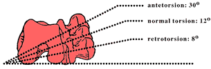
For purposes of clarity, the following illustrates the superimposition of normal torsion, retrotorsion, and antetorsion.
Remember: when speaking of the TORSION angles, the head and neck of the femur are measured relative to the condyles of the femur. When speaking of the VERSION angles, the head and neck are measured relative to the frontal plane of the body. To keep the terminology and concepts without confusion, I suggest the following crutch. If Antetorsion causes and internal rotation of the limb then Retrotorsion will cause an external rotation of the limb. If Antetorson causes an internal deviation and Anteversion will cause the opposite i.e., external rotation.
In the normal adult, the "Torsion" angle equals the "Version" angle. "Torsion"="Version"=12 degrees.
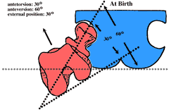
At birth, the torsion within the femur and the position of the femur relative to the frontal plane of the body are markedly different from adult values. Examination of the newborn should demonstrate an externally rotated limb by approximately 30 degrees. Antetorsion=30 degrees
The limb is externally rotated because the increase in anteversion is 30 degrees greater than the increase in antetorsion. From birth to adulthood anteversion decreases from 60 degrees to 12 degrees for a difference of 48 degrees. Therefore the net effect of this decrease is a positional change of 48 degrees resulting in a less externally deviated limb. At the same time, Antetorsion reduces from 30 degrees to 12 degrees for a difference of 18 degrees. The net effect of this decrease in antetorsion is a structural change resulting is less internal deviation of the limb. These changes usually occur in the first six years of life but may be delayed until adolescence.
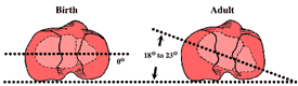
Statistically, 95% of abnormal torsion of the femur reduces by adolescence. Between age 6 and adolescence, the presence of an in-toe gait secondary to an internal torsion may indicate a torsional abnormality that may proceed into adulthood or a delay in development. While the clinician may have to wait for adolescence to verify spontaneous reduction, a good clue may be found in the families of the child's parents. The presence of an in-toe gait within the child's adult family is highly suggestive of a true torsional abnormality (and the limb will not derotate with time) while the absence of an adult with an in-toe gait within the child's family indicates delayed development and probable spontaneous resolution. At birth there is no tibial (malleolar) torsion but soon develops with weight bearing. Normal adult value for malleolar torsion is 18 to 23 degrees. A decrease in this value leads to an adducted gait while and increase in this value leads to an abducted gait. The position of the foot in the transverse plane (abducted or adducted) is not readily altered by gait plates (see later).
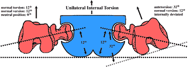
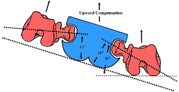
THE CLINICAL PICTURE The following description of the clinical presentation is divided into two parts. The first describes the most common responses for the foot i.e., downward compensations while the latter will describe the uncommon but intriguing upward compensations that are associated with internal torsions of the femur. With either a retrotorsion or an anteversion, the limb will be externally deviated and the neutral position of the hip rotations will be external. The knee and foot will function externally and an increase in the angle of gait will be noted clinically. With external torsions/positions there is a retrograde pronatory force on the foot that can be easily demonstrated. If you stand up, abduct your feet significantly, and lean forward and (voila!) your feet will pronate, illustrating the retrograde pronatory force. Abducted gait secondary to external torsion/position of the femur respond well to foot orthoses. A standard device will usually reduce the angle of gait to normal limits but on occasion a gait plate may be necessary. With either an antetorsion or a retroversion, the limb will function internally deviated and the neutral postions of the range of motion of the hip will be internal. In gait, the knee is readily noted to be internally deviated. At this point the patient has three choices: (a) let the limb and foot be internally deviated (adducted foot), (b) use active muscle contraction to externally rotate the hip or (c) contract peroneus brevis muscle to abduct the foot. In the uncomplicated scenario the foot will be adducted and the foot will have a retrograde supinatory force. (Stand up, adduct your feet and lean forward and (voila!) the foot supinates.) The problem with an adducted gait is primarily cosmesis, not function; the foot does well, all things considered. The pigeon toe gait is usually unacceptable to adults and children and compensations result. With children, either the parents or the child realize the gait is peculiar and attempt to alter the angle of gait. The limb could be rotated externally at the hip but this takes too much muscle effort. It is much easier and more efficient to contract the peroneus brevis muscle to abduct the foot and thereby produce a more normal angle of gait. The peroneus brevis is normally a stance phase muscle that is recruited to fire during the swing phase of gait to abduct the foot. Unfortunately, the foot is abducted (pronated) in swing, abducted and pronated during the entire stance phase of gait from heel contact through toe off. This is open kinetic chain pronation of the foot. The foot never resupinates and is generally a very pathological , symptomatic and subluxed foot. Shoe wear is pathonomonic: excessive wear on the medial aspect of the heel and the medial aspect of the sole of the forefoot and the footgear is generally short lived. Thus with internal deviations, two extremes may be seen: a child/adult with an adducted gait with a retrograde supinatory force on the foot or a relatively normal angle of gait with a subluxed foot secondary to open kinetic chain contraction of the peroneus brevis muscle. When using orthotics to treat a flat foot secondary to an internal deviation of the limb cosmesis is once again a concern. Control of the pronated foot with regular orthotics will lead to an adducted gait. Both adults and children should be informed of this consequence. Gait Plates may be employed but the response is not uniform. It is generally true that Gait Plates positively alter the angle of gait for torsional and positional abnormalities of the femur but usually do not alter the angle of gait for torsional abnormalities of the tibia (increased or decreased malleolar torsion). Upward compensation for internal torsions are intriguing. As mentioned earlier, downward compensation for an internal femoral torsion (antetorsion) the extremes of foot function may be represented by an adducted gait (with relatively good foot function) or a very flat foot (open kinetic chain pronation) with a relatively normal angle of gait. Upward compensations are usually associated with a unilateral internal torsions or assymmetrical bilateral internal torsions, i.e., one extremity is more severely affected than the opposite extremity. Illustrated below are unilateral internal torsions of the femur on the right extremity. The first illustration shows a pelvis, its plane of progression (straight ahead), a normal left extremity and an internally deviated right extremity. The right foot may be adducted or it may be subluxed secondary to open kinetic chain pronation. In the second illustration (same scenario of internal torsion of the right limb only) the patient has learned to lead with the opposite hip (upward compensation). Although the pelvis is oblique, the plane of progression of the body remains straight ahead. Note that the right limb is parallel to the plane of progression thereby decreasing the necessity for right pedal compensation, although it may still occur but to a lesser extent. Most interesting in the end result to the left extremity. Now the left limb is internal relative to the plane of progression but is in normal alignment relative to the pelvis. Note that the foot which is opposite to the side of the deformity is internally deviated (adducted) relative to the plane of progression.
Clinically, the left foot may be adducted and relatively stable. The other extreme is that the left foot could be subluxed secondary to open kinetic chain pronation (attempting to improve the angle of gait). In this last example the flat foot is on the opposite limb to the major deformity. Hip leading (upward compensation) will also occur with bilateral but asymmetrical internal torsions, or in other words one limb is more severely affected with internal torsion. In these cases the hip leading is always on the opposite side to the major deformity. Hip leading is readily observable in gait. At heel contact, the affected side always displaces further forward than the opposite pelvis at heel contact. The obliquity of the pelvis relative to the plane of progression can be observed when looking at a complete gait cycle. Remember, the pelvis is still oscillating in the transverse plane but functioning obliquely to the plane of progression. |