 Case of
the week - 15/05/98
 Case of
the week - 15/05/98

Manual muscle test (MRC grading)
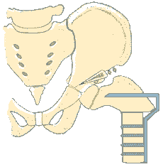
Hip abductors: left 4 right 2
Knee flexors: left 4 right 4
Knee extensors: left 4 right 5
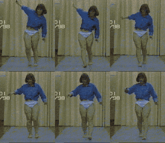
Movies (PC users get the QuickTime plug-ins here - right mouse-click "Save Link as..." - Windows 95/NT and 3.1)
The movies will open in new browser windows so you can view them simultaneously
with analysis.
|
|
frontal | |
| movie format | QuickTime | QuickTime |
| left side | front view | |
| right side | rear view |
Poly-Electromyography (Noraxon) ... Right-side..... Left-side
Note: on the left side the gait cycle is, unusually, from toe off to toe off, and the blue bars indicating normal activity are incorrect - she actually shows normal activity from initial contact to the next inital contact (marked by the vertical line). EMG of the right side is, as usual, from initial contact to inital contact and the blue bars in this case correctly represent normal activity.
... points for discussion:
 What people said...
Case supplied by Mag.Andreas
Kranzl and Dr. Andreas
Kopf
from Univ. Clinic for Physical Medicine & Rehabiliation, Vienna
General Hospital
Email your comments to [n/a]
Maintained by DDr.
Chris Kirtley, Andreas
Kranzl & Dr. Andreas
Kopf
Last modified on Wednesday, 27-Feb. 1998.