 Teach-in
'99 : Optical versus Electromagnetic Motion Analysis
Teach-in
'99 : Optical versus Electromagnetic Motion Analysis
by Raymond Lee & Chris
Kirtley, The Hong Kong Polytechnic University
This experiment compared the output of Vicon video-based
motion analysis with an electromagnetic tracking device (Polhemus Tracker).
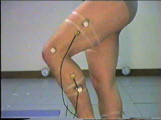
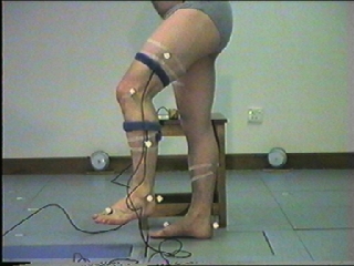
As shown in the figures, the standard Vicon Clinical Manager marker
set was used and the two Tracker sensors were attached (using double-sided
adhesive tape and tight velcro straps) to the thigh and shank segments.
The subject then performed a series of flexion-extension movements of the
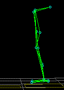
knee, as seen in the animation.
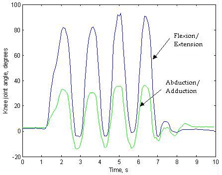
The VCM analysis is shown below:
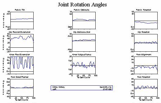
The sagittal plane flexion-extension of the knee is well
recorded. However, note two things:
-
there is a flexion offset at the knee, caused by the the
knee marker being placed too anteriorly
-
there is a small abduction/adduction (sometimes erroneously
called "varus/valgus", terms which apply only to static deformities) artifact,
caused by the thigh wand being slightly rotated with respect to the femur
The Tracker output shows a similar artifact:
In this case, there is a slight hyperextension offset,
and an abduction/adduction of around 40 degrees peak to peak.
With the Tracker, it is quite a straightforward
matter to realign the measurement axes of the sensor, and this was done
in order to bring the two sensors into alignment defined from a static
trial. After this correction, the following results were achieved:

\
Note that the artifact is now much reduced (about 20
degrees peak-to-peak), and is now of similar magnitude to the Vicon
data.
Questions
-
What is responsible for the residual artifact?
-
How could it be further reduced?
-
Is there any fundamental difference or limitation in optical
motion analysis versus electromagnetic tracking?
Email your answers to [n/a] 
 What
we said on the CGA list
What
we said on the CGA list
 Back to Teach-in page