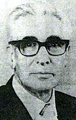
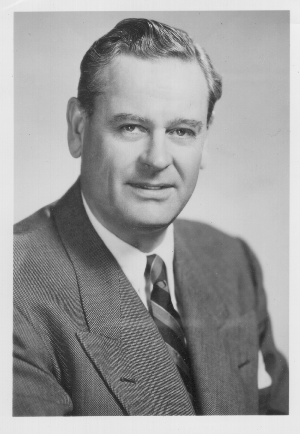
 Verne Inman
was an exceptional person in an important position at a pivotal time. Not
only did his studies of the biomechanics of locomotion establish the field,
they are classics in the application of engineering expertise to clinical
problems through basic research.
He was born in San Jose, California in 1905 and attended the University of California at Berkeley as an undergraduate. While in medical school, he worked as an assistant in anatomy and earned an M.A. degree for studies of electrical stimulation of cutaneous nerves in humans.
Upon completing his medical studies he commenced a Ph.D in Anatomy. His specific interest focused on nerve conduction in the giant axon of invertebrates, which fitted well with the department's emphasis on physiology. While the electrical equipment he needed to conduct this project was available from his prior research, he was unable to obtain suitable specimens and had to abandon the project. Although disappointed, he chose to study the differential development of the human fetal cranium, and with that work obtained his Ph.D in 1934, being the last doctoral student at Berkeley or San Francisco to offer a thesis in gross anatomy. An internship and residency in orthopaedics followed, and by 1940, Verne Inman had become a clinical instructor in orthopaedic surgery as well as an instructor in anatomy, and had published articles on the anatomy and pathology of the intervertebral disc.
As World War II neared its end, he published an important paper on the biomechanics of the shoulder. Shortly thereafter, at the urging of the National Research Council, the Surgeon General's Office (and, later on, the Veteran's Administration) he began developing a research program directed at the physical problems of the tens of thousands of amputees who were returning from the war. The upper-extremity research program, which might have been a logical extension of his shoulder research, was established in Southern California to be near the aerospace industry which had the flexible cables necessary for such research. The lower-extremity research program, in which he was one of two major leaders, began in Northern California as the Biomechanics Laboratory. It was located on both the Berkeley campus in collaboration with faculty from the Department of Engineering, and on the San Francisco campus, initially using space in the Department of Anatomy.
It was soon evident that basic research was needed if intelligent design
changes were to be made in the unsatisfactory prosthetic devices of the
time. Here, then, was the opportunity for him to integrate his interests
in mechanical and electronic devices, since
elucidation of the basic biomechanics of locomotion required study
of not only the dynamics of body motion, but also analysis of the phasic
electrical activity of the appropriate muscles. As the scope of the project
broadened to include problems with muscle physiology, pain, and skin, the
Biomechanics Laboratory became the first departmentally based interdisciplinary
research group on the San Francisco campus. The collaboration between the
basic sciences and the Department of Orthopaedic Surgery established high
standards for such worthwhile interactions. The goals of basic research
as well as their clinical applications were met by the diverse group of
specialists who participated in the project. International fame and major
publications in the areas of human locomotion, the mechanisms of pain,
and rational prosthetic design were the result.
Verne's role was often to reframe a question, simplify the approach
to a problem, or outline a general research strategy, and then step aside
to let the specialist, researcher, or student proceed. He had a consuming
curiosity and thoroughly enjoyed the challenge of problems while maintaining
a resolute skepticism when confronted with superficial or pat answers.
He saw
issues broadly, and sought solutions in unexpected sites. In one instance,
for example, his dissection of a bear's foot provided insight into the
anatomy of the human plantigrade foot. The elegance of nature revealed
by his research gave Verne inordinate pleasure. He also enjoyed working
skillfully with his hands; it was not uncommon to find him in the laboratory
or shop on weekends or late at night, constructing a beautifully detailed
working model with which to demonstrate some fundamental principle of human
biomechanics.
Retirement in 1973 did not change Verne's approach to life. His time and energies simply turned to the problems of the family "ranch" in San Jose. There, he designed a new means of clipping large hedges, and he cultivated unusual plants and fruit trees while remaining a consultant to the Biomechanics Laboratory. He met with his editorial staff to finalize the manuscript of his definitive treatise Human Locomotion just three weeks before his death after a brief illness at age 74.
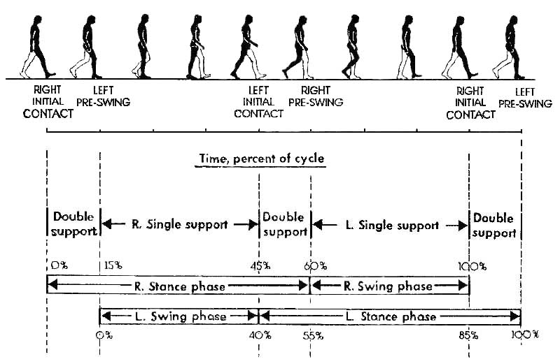
Though published over 40 years ago, this list of gait determinants still guides many researchers and clinicians' thinking about human gait. The authors began by assuming that a gait pattern is most efficient when it minimizes vertical and lateral excursions in the body's center of gravity (COG). They identified those features of the movement pattern that miminize these COG excursions. They suggested that these features determine whether a movement pattern is normal or pathological.
Inman, VT, Ralston, HJ, and Todd, Frank (1981). Human Walking.Williams
and Wilkins, Baltimore/London.
Saunders, J.B., Inman, V.T., & Eberhart, H.D. (1953). The major
determinants in normal and pathological gait. Journal of Bone and Joint
Surgery, 35A, 543-558.
Inman, V.T. (1966). Human locomotion. Canadian Medical Association
Journal, 94, 1047.
Mann, Roger, and Verne T. Inman 1964. Phasic activity of intrinsic
muscles of the foot. Journal of Bone and Joint Surgery
46A(3):469-481.
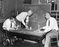 Dr. Verne Inman (center), Professor Howard Eberhart (right), and __ Henderson
(in wheelchair) in the Biomechanics Laboratory at Berkely.
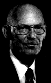
Prof. Eberhart was admitted to the National Academy of Engineering in
1977 "for pioneering studies of human locomotion, application of structural
engineering to prosthetic devices, and leadership of interdisciplinary
engineering research".
Saunders came to the University of California in 1931 as an anatomy
professor, was chair of the department from 1938 to 1956. He conducted
research with LeRoy C. Abbott of the Department of Orthopedic Surgery,
and Verne T. Inman of the
Biomechanics Laboratory. With them and other colleagues he studied
the normal and diseased spinal column, nerve injury and
regeneration, the mechanics of the joints of the upper and lower limbs,
and the nature of normal and pathological gait. He published studies on
congenital abnormalities of the duodenum, and on the development of the
genitourinary tract. Later, he made important observations on the formation
of the ventricles and the aorta.
On the occasion of the 400th anniversary of the publication of the great anatomical work, De Humani Corporis Fabrici, by Andreas Vesalius in 1543, Saunders published a paper with Leroy Crummer on Loys Vasse, an contemporary of Vesalius. He continued to write several papers on aspects of the life and times of Vesalius and continued to write and publish on medical history until his death at age eighty-eight.
Saunders, J.B., Inman, V.T., & Eberhart, H.D. (1953). The major
determinants in normal and pathological gait. Journal of Bone and Joint
Surgery, 35A, 543-558.
 Boris Bresler
(1919-2000)
Boris Bresler
(1919-2000)Energy and power in the legs of above-knee amputees during normal level
walking, B. Bresler, C,W. Radcliffe, and F.R. Berry. Berkeley, Lower-Extremity
Amputee Research-Project, Institute of Engineering Research, University
of California, 1957. Radcliffe, C. W. (Charles W.) Technical report (University
of California, Berkeley. Biomechanics Laboratory); 31.
Bresler, B. and Frankel, J. (1950). The forces and moments in the leg
during level walking. Transactions of the ASME: 27-36.
Due to political reasons, his1947 book, Coordination and Regulation of Movements, was not translated until 1967. Bernstein challenged the view held in McGraw's and Gesell's time of a hierarchical system within the body whereby commands for movement were issued by the brain. He posited that performance of any kind of movement results from an infinite variety of possible combinations, or degrees of freedom, of neuromuscular and skeletal elements. The system should, therefore, be considered as self-organizing, with body elements coordinated, or assembled, in response to specific tasks. Motor development was dependent not on brain maturation, but adaptations to constraints of the body (changes in the growing infant's body mass and proportions) and to exogenous conditions (gravity, surface, specific tasks to be performed).
The analysis of coordinated movements became the study of biomechanics.
This term, coined by Bernstein, describes the application of mechanical
principles and methods to biological systems. These involve two areas:
kinematics, or time-space forms of movement such as muscle activation and
joint movement; and dynamics, the physical causes of movements such as
inertial or centripetal forces, power (acceleration) and torque (rotation).
The methods of analysis include electrographic recordings of joint angular
velocity and muscle activation, and free body diagrams showing body segments
and forces acting on them
Space requirements of the seated operator. Technical Report USAF,
WADC TR-55-159 (AD 87 892), Wright Air Development Center, Wright-Patterson
Air Force Base, Ohio, 1955.
Wilfrid Taylor Dempster. The anthropometry of body action. Annals New York Academy of Sciences, 63:559{585, 1956.
Rudolfs Drillis and Renato Contini. Body segment parameters. Technical
Report 1166-
03, New York University, School of Engineering and Science, Research
Division, New York under contract
with Office of Vocational Rehabilitation, Department of Health, Education
and Welfare, September 1966.
Charles E. Clauser, John T. McConville, and J. W. Young. Weight, volume,
and center of
mass of segments of the human body. Technical Report AMRL-TR-69-70
(AD-710 622), Aerospace Medical
Research Laboratory, Aerospace Medical Division, Air Force Systems
Command, Wright-Patterson Air Force
Base, Ohio, August 1969.
Based on these calculations the three principal mass moments of inertia
of each body segment
were estimated.
Some of the results of this study of cadavers were compared to those
obtained, by Santschi and
coworkers, on living subjects and it was concluded that a sat- is factory
level of agreement exists.
It was also concluded that the principal mass moments of inertia of
body segments correlates
well with total body mass and (especially) with segment volume.
R. F. Chandler, C. E. Clauser, J. T. McConville, H. M. Reynolds, and
J. W. Young.
Investigation of inertial properties of the human body. Technical Report
DOT HS-801 430, Aerospace Medical
Research Laboratory, Wright-Patterson Air Force Base, OH, March 1975.
Exoskeletal goniometer examination of lower limb motions during walking
at different speeds.
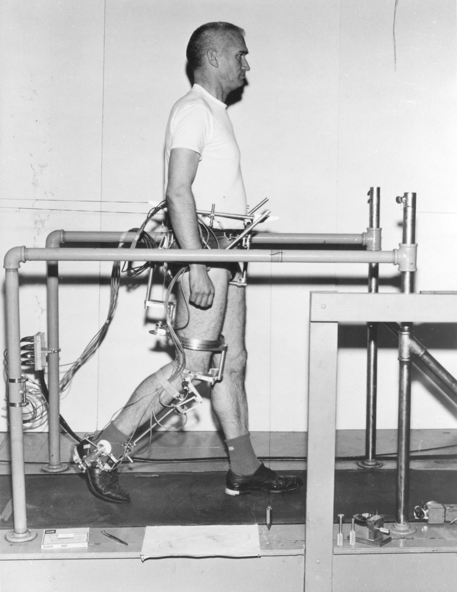
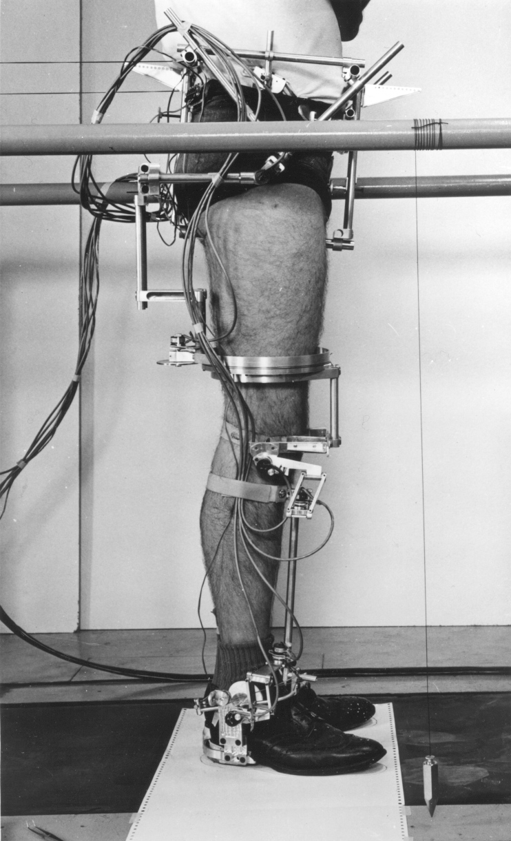

The pelvic frame was attached with eight contacts, over the ASISs, iliac crests, pubis, sacrum, and ischial tuberosities. The hip joint was an Euler sequence - flexion, abduction, axial rotation - that was aligned to the anatomic hip joint. Knee and ankle joints were measured with parallelogram linkages that did not depend on precise alignment with the anatomic axes for accurate angle measurements. Measurements were conducted on a treadmill to allow averaging of multiple cycles. Pelvic motions, both linear and angular, were recorded by parallel strings attached to displacement transducers mounted to the treadmill frame. Potentiometers provided electrical outputs which were recorded onto an FM modulated instrumentation tape recorder. Replay from the recorder was converted to digital format on a DEC PDP-7 computer (18 bit).
Lamoreux, LW, 1971, Kinematic Measurements in the Study of Human Walking, Bul. Prosth. Res. BPR 10-15
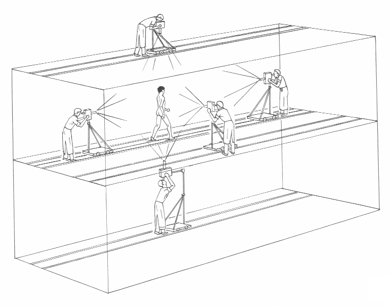 The Glass Cage
Several novel tools are described in the book, including the "glass
cage" and "simplified glass cage".

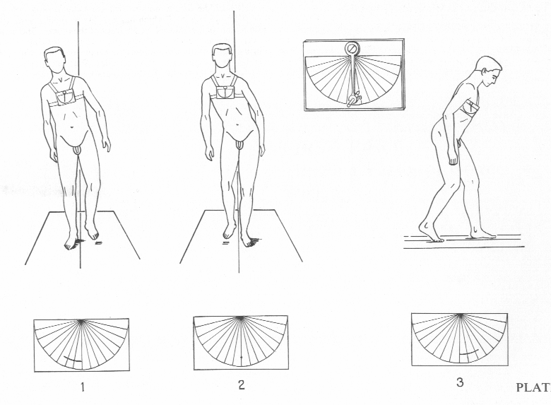
The orthopedist is encouraged to observe the gait in 3D and use a hand-held
sighting goniometer and "claudico-oscillometer" to quantify pathological
angles.

Observing gait in 3D with the simplfied Glass Cage
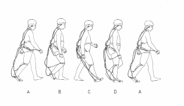
 Saussez equipment

Canadian anatomist, earned his medical degree from the University of Toronto in 1945 where he then he joined the staff and achieved the rank of Professor. Along the way he also pursued clinical research at the Hospital for Sick Children and McMaster Univ. His work with polio patients using electromyography to study muscle control mechanisms revolutionized the use of electronic and mechanical devices for people with neurological and orthopedic impairments. Introduced fine-wire electrodes that were more comfortable than needles and could be used longer. In the 1960s, he used electromyographic biofeedback to train patients to regain control muscle functions that were thought to be permanently lost. It is because of these efforts that he is referred to as the "Father of EMG Biofeedback Therapy". Founded International Society of Electrophysiological Kinesiology, ISEK, in 1965 to create standards for EMG usage and reporting. This society continues today as a multidisciplinary organization dedicated to studying human movement and the neuromuscular system.
Dr. John Basmajian received his MD degree in 1945 from the University of Toronto. He intended to specialize in Orthopaedic Surgery but for health reasons specialized in Anatomy.
Muscles Alive was the first collection of studies that used technology to study muscle behavior during voluntary activity. This book sparked the imaginations of countless students and practitioners of Health Sciences, Medicine, and Engineering to explore the workings of muscles and, as he put it "their functions revealed by Electromyography". His passion and tireless curiosity for understanding human movement in the normal and dysfunctional states brought forth more than two dozen books and nearly 400 scientific papers. He was awarded the highest civilian honor as an Officer of the Order of Canada (OC).
Basmajian, J. V. (1978). Muscles alive: Their functions
revealed by electromyography. 4th ed Baltimore: Williams
and Wilkins.
On psychological aspects of backaches, Time 14 Jul 80
Wolf SL (1997) The first Basmajian lecture. Reflections on John V. Basmajian:
Anatomist, Electromyographer, Scientist.J Electromyogr Kinesiol. 7(4):213-219.