 Emst
Heinrich (1795-1878), Wilhelm Eduard (1804-1891) & Eduard Friedrick
Wilhelm Weber (1806-1871)
Emst
Heinrich (1795-1878), Wilhelm Eduard (1804-1891) & Eduard Friedrick
Wilhelm Weber (1806-1871)Die Mechanik Der Menschlichen Gerverzeuge (1836)
E. Harless (1860). The static moments of human limbs (in German). Treatises of the Math.-Phys. Class of the Royal Acad. of Sc. of Bavaria, 8:69{ 96 and 257{294, 1860.
E. Harless. The static moments of the component masses of the human body. Trans. of the Math-Phys., Royal Bavarian Acd. of Sci., 8(1,2):69{96 and 257{294, 1860. Unpublished English Translation, Wright-Patterson Air Force Base, Ohio.
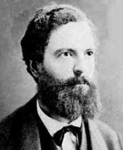
 Étienne-Jules
Marey (1830-1904)
Étienne-Jules
Marey (1830-1904) 


All movement is the product of two factors: time and space; to know
the movement of a body is to know the series of positions that it has occupied
in space in a series of successive moments.
Introduction to La Methode Graphique dans les Sciences Experimentales
1878
Marey’s study of walking and research on muscular forces, as well as his work on physical education and gymnastics in schools, gave him the chance in 1882 to request the French Ministry of War and Ministry of Public Education to finance the work and running costs of his laboratory, the Station Physiologique. Marey defined the project of the Station as: "to determine the series of actions which are created in human locomotion in its various types [...], to measure the effort expended at each moment in the different actions of locomotion, in order to seek the most favourable conditions for utilisation of that effort." His research allowed the establishment of rules governing the manoeuvres of soldiers, the physical exercises of young people, and the operations carried out by workers: in short, the best possible use of the muscular effort of the human being.
 To assist in
these studies, the Ministry of War put a number of infantry soldiers, who
had volunteered to take part in experiments, at the disposal of Marey and
his assistant Demenÿ. One Lieutenant Andriveau was delegated to supervise
the work of the two scientists, and to report their results to the Army
at regular intervals. Soon Andriveau and Demenÿ were working on their
own; Marey was in Naples, from where he followed the work of the Station
at a distance, receiving reports and offering his advice.
To assist in
these studies, the Ministry of War put a number of infantry soldiers, who
had volunteered to take part in experiments, at the disposal of Marey and
his assistant Demenÿ. One Lieutenant Andriveau was delegated to supervise
the work of the two scientists, and to report their results to the Army
at regular intervals. Soon Andriveau and Demenÿ were working on their
own; Marey was in Naples, from where he followed the work of the Station
at a distance, receiving reports and offering his advice.
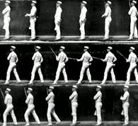 Demenÿ and
Andriveau worked tirelessly, even in deepest winter. They used both the
equipment of the graphical method (such as the Odographe and Dynamographe)
and glass-plate and celluloid film chronophotography to record the efforts
of the soldiers. The soldiers were very carefully measured and weighed,
and had to perform all sorts of physical exercises, such as running or
walking around the track of the Station to the rhythm of an electric beater.
They were also required to run carrying loads of 30 to 40 kilos (approximately
65 to 90 pounds), with the aim of measuring their fatigue to allow organisation
of battalions on the basis of the strength of their individual members.
Demenÿ and
Andriveau worked tirelessly, even in deepest winter. They used both the
equipment of the graphical method (such as the Odographe and Dynamographe)
and glass-plate and celluloid film chronophotography to record the efforts
of the soldiers. The soldiers were very carefully measured and weighed,
and had to perform all sorts of physical exercises, such as running or
walking around the track of the Station to the rhythm of an electric beater.
They were also required to run carrying loads of 30 to 40 kilos (approximately
65 to 90 pounds), with the aim of measuring their fatigue to allow organisation
of battalions on the basis of the strength of their individual members.
 Economisation
of human muscular effort was a topic which fascinated Demenÿ, who
had been working on the development of physical education for several years.
The soldiers – when they were not athletes from his rational gymnastics
society – gave him the opportunity to prove his theories of gymnastics.
He filmed men performing high and long jumps, sprinting, lifting objects,
pulling on ropes, and so on. His theory was that well-developed athletes
would subconsciously use those positions and movements which were least
damaging to the body, and these could then be recommended to the military.
Economisation
of human muscular effort was a topic which fascinated Demenÿ, who
had been working on the development of physical education for several years.
The soldiers – when they were not athletes from his rational gymnastics
society – gave him the opportunity to prove his theories of gymnastics.
He filmed men performing high and long jumps, sprinting, lifting objects,
pulling on ropes, and so on. His theory was that well-developed athletes
would subconsciously use those positions and movements which were least
damaging to the body, and these could then be recommended to the military.
 Marey and Demenÿ
also turned their attention to the study of workers, on the basis that
filming the actions of a blacksmith, a house painter, or a sawyer, might
be used to help training for these trades. All that would be needed to
become rapidly qualified, they thought, would be to carefully observe the
positions and actions of a worker, then repeat them as faithfully as possible.
Marey and Demenÿ
also turned their attention to the study of workers, on the basis that
filming the actions of a blacksmith, a house painter, or a sawyer, might
be used to help training for these trades. All that would be needed to
become rapidly qualified, they thought, would be to carefully observe the
positions and actions of a worker, then repeat them as faithfully as possible.
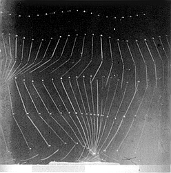
By 1867 Marey had begun investigating the external motion and
movement of bodies. Using ingenious recording devices he
studied the gaits of horses and humans, and examined how
birds and insects achieved flight. While Marey's graphic method
continued to produce significant results, it had several distinct
disadvantages when applied to the study of bodies in motion. It
required a physical connection between the thing being
measured and the recording device itself, a connection which
often times constrained the natural movements being studied. It
was also unable to record the changing positions of a body (or
limb) moving through space. While Marey would continue to
use the graphic method in future experiments, after seeing
Eadweard Muybridge's photographs of horses in motion, the
possibility of using photography to extend the range of his
investigations was too tempting to pass up.
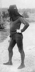

Marey's man in black velvet became the inspiration for Marcel Duchamp's
Nude
Descending a Staircase.


 sphygmograph,
used to measure the pulse. One end of a lever rested on the veins in the
wrist, while a stylus on the other end inscribed the fluctuations of the
heart onto a carbon-blacked strip of paper moving at a uniform speed.
sphygmograph,
used to measure the pulse. One end of a lever rested on the veins in the
wrist, while a stylus on the other end inscribed the fluctuations of the
heart onto a carbon-blacked strip of paper moving at a uniform speed.
 'Fusil Photographique' or Photographic Gun
![]()

Honorary-degree ceremony outside the Old Schools, Cambridge (1898)
from left to right: E.J. Marey, A. Dohrn, C. Golgi, E.H.P.A. Haeckel, A.A.W.
Hubrecht, W.F. Kuehne, H.P. Bowditch, Hugo Kronecker and H. Kronecker (Wellcome
Museum, London).
Braun, Marta Picturing Time: The Work of Etienne-Jules Marey (1830-1904). xxii, 450 p., 270 halftones, 65 line drawings. 8-1/4 x 9-1/2 1992, Paper 0-226-07175-8
Musee de Marey, Beaune in Burgundy, France.
Marey EJ (1885) La Methode Graphique, Paris
Marey EJ (1895) Movement, London
Tufte ER (1983) The Visual Display of Quantitative Information, Graphics Press, Cheshire, Connecticut.

 Zoopraxiscope
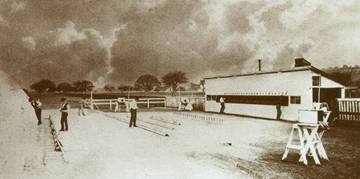 Experiments


Braune and Fischer made a very careful study of mass, volume and center
of mass of three adult
male cadavers and their body segments. The cadavers were close to the
average build of German
soldiers of that period and they were all dead from suicides. To avoid
fluid loss etc. the cadavers
were kept frozen throughout the study. The center of mass of each body
segment were not
estimated by the use of balance plates, as in the previously described
studies, but by driving thin
rods into the tissue and hanging the body segment from three axes.
The intersection of three
externally fixed planes, e.g. vertically through each of the axes,
formed on the segment,
corresponds to the center of mass.
This study was so thorough that it uncritically was used as a standard
for more than half a
century, despite the pronounced differences in and between populations.
Braune and Fischer also
introduced the use of regression equations for estimation of body segment
parameters, based on
the length and mass of body segments. 1894 - Meeh Meeh pointed out,
that the results obtained from cadavers should be supplemented with
data from living subjects.
To estimate the volume of body segments, each segment was immersed
in water up to the joint
and the amount of water displaced thereby was measured. This method
was found to be inexact
and each measurement was therefore repeated several times and averaged.
Using the densities found
by Harless, Meeh was able to estimate the absolute and relative mass
of each body
segment from its volume and to make a series of graphs to illustrate
the growth of the body and
its segments as a function of age. This was the first serious attempt
to describe the changes in
mass of body segments during growth.
Interestingly, Braune was the son-in-law of Emst HeinrichWeber.
Braüne W. and Fischer O. (orig. published 1895-1904.) The Human
Gait. Springer-Verlag, Berlin, 1987.
Fischer O (1899) Abh K Sächs Ges. Wiss. Math.-Phys. Classe 25:
1
Fischer O (1901) Abh K Sächs Ges. Wiss. Math.-Phys. Classe 26:
471
Fischer O (1904) Abh K Sächs Ges. Wiss. Math.-Phys. Classe 28:
533
Fischer O (1906) Theoretische Grundlagen für eine Mechanik der
lebenden Körper. Leipzig.
Trendelenberg' s name is associated with two clinical signs. The first, described in 1890, identified saphenofemoral incompetence in patients with varicose veins. The second, described in 1895, confirms shortening of the leg due to an ununited fracture of the femur or dislocation of the hip joint. The operative position named after Trendelenberg consists of placing the patient in a 45 degree 'head down' inclination, useful in reducing the venous pressure for varicose vein surgery or to maintain the intestine out of the pelvis for gynaecological procedures. The position was first described by Willy Meyer who had been a student under Trendelenberg at Bonn in 1881.
Trendelenberg was a great practical surgeon who was keenly interested in the history of surgery. His doctoral thesis De Veterum Indorum Chirurgiais discussed ancient Indian surgery. The closing years of his life were spent in Nikolassea, Germany. He died from carcinoma of the lower jaw at the age of 81 years.
The original paper on Trendelenberg Gait (click
each page to see translation by Jean Lieberman & Larry Lamoreux):
![]()
![]()
![]()
![]()
 Born
September 17, 1806, Boulogne-sur-Mer of a family of fishermen, traders
and sea captains who had resided in the harbour city Boulogne-sur-Mer in
Northern France since the first half of the 18th century. He was predestined
for a career at seas, as his father was the commander Jean Duchenne who
had been a ships captain during the Napoleonic wars, and expected his son
to follow in his keel waters. However, it was his love of science
that prevailed. Duchenne went to a local college at Douai, where he received
his baccalauréat at the age of 19. From 1827, aged twenty-one, he
studied medicine under teachers like René-Théophile-Hyacinthe
Laënnec (1781-1826), Baron Guillaume Dupuytren (1777-1835), François
Magendie (1783-1855), and Léon Cruveilhier (1791-1874). He graduated
in medicine in Paris in 1831 and, probably influenced by Dupuytren, presented
his Thèse de médecine, a monograph on burns. According to
Ernest-Charles Lasègue (1816-1883), and Isidor Straus (1845-1896)
he was ”of medium height, thickset, active in movement and slow in speech.
Born
September 17, 1806, Boulogne-sur-Mer of a family of fishermen, traders
and sea captains who had resided in the harbour city Boulogne-sur-Mer in
Northern France since the first half of the 18th century. He was predestined
for a career at seas, as his father was the commander Jean Duchenne who
had been a ships captain during the Napoleonic wars, and expected his son
to follow in his keel waters. However, it was his love of science
that prevailed. Duchenne went to a local college at Douai, where he received
his baccalauréat at the age of 19. From 1827, aged twenty-one, he
studied medicine under teachers like René-Théophile-Hyacinthe
Laënnec (1781-1826), Baron Guillaume Dupuytren (1777-1835), François
Magendie (1783-1855), and Léon Cruveilhier (1791-1874). He graduated
in medicine in Paris in 1831 and, probably influenced by Dupuytren, presented
his Thèse de médecine, a monograph on burns. According to
Ernest-Charles Lasègue (1816-1883), and Isidor Straus (1845-1896)
he was ”of medium height, thickset, active in movement and slow in speech.
Duchenne had an undistinguished undergraduate career and as he failed to obtain an academic post, and because of his father’s death he returned to Boulogne where he practised medicine for ten years. Duchenne married in 1831, but his wife died in childbirth already to years later - of puerperal fever. Duchenne’s mother in law spread rumours that the death of his wife was caused by the fact that only he was present at the delivery, and after this he was separated from his only son by his wife’s family. As a consequence of this his patients the following years avoided his previously blossoming practice. After a new short and unhappy marriage he returned to Paris penniless in 1842 in order to pursue his ambition of doing medical research. His interest was particularly directed towards electropuncture, which had recently been invented by Magendie and Jean-Baptiste Sarlandière (1787-1838).
In Paris Duchenne met with a rather cool reception, being ridiculed for his provincial accent and his course manners. He was known under the name of Duchenne de Boulogne to avoid confusion with Édouard Adolphe Duchesne (1894-1869), a fashionable society physician. Having arrived alone and without funds, he set to work in charity clinics and hospitals, and gained his livelihood from private practice, which presumably became adequate to supply his limited needs. He seemed to live only for his patients and his scholarship, pursuing his clinical neurological studies in a very unorthodox but effective fashion. He was described perhaps somewhat romantically as a strange mariner-like figure that every morning used to haunt one or two major Parisian teaching hospitals in order to study the most interesting cases, then to make them an object of his electrotherapeutic detailed studies. Duchenne was a lonely figure at the wards of the Paris hospitals, mocked by the interns and rebuffed by the senior medical staff, whom he called monarchs of the ward. However, he demonstrated an enormous personal courage and a singleness of purpose, and soon obtained a reputation as an outstanding neurologist and thus achieved some degree of academic recognition. However unambitious Duchenne may have been, he was frequently involved in disputes concerning forms of diseases that he had discovered, and he was never short of enemies and protagonists.
Duchenne built his own machine for neuromuscular stimulation and developed the technique of using surface electrodes. In 1861 in the second edition of his book, Paraplégie Hypertrophique de l’enfance de cause cérébrale, Duchenne described a boy with the form of muscular dystrophy that now bears his name. A keen photographer, he depicted his patient a year later in his Album de photographies pathologiques. He discovered that external electrical stimulation could cause muscle movements and initially used this as a form of therapy, but then appreciated its possibilities as a diagnostic method. He developed the technique of using surface electrodes. Previous electric stimulation had caused a lot of tissue damages. The power source, portable in order to be usable at bedside, consisted of a coal-zinc battery. Exactly how the stimulation was performed is not known, but it resulted in either repeated contractions or tetanus. He employed this technique to analyse the mechanism of facial expression, the photographsof which were published by Darwin reproduced in The Expression of the Emotions in Man and Animals. He distinguished between indirect stimulation via the nerve» and direct stimulation of the muscle. The first results were described in a report to the Académie de Médecine in Paris in 1848.
In the first two editions of De l'Electrisation localisée Duchenne renders exact clinical descriptions of the natural course of polio. By a series of observations and deductions he proved that acute poliomyelitis (previously called ”paralysie essentielle de l’enfance,” localisation unknown) was a disease of motor nerve cells in the spinal cord. He suggested - without having any histological basis - that the profound paralysis of poliomyelitis must be due to a lesion that he located in the anterior horn cells of the spinal cord. This hypothesis proved correct when André Victor Cornil (1837-1898) in 1863 and Charcot in 1870 demonstrated histological changes.
During the later part of his life Duchenne was a happy man, particularly after he had had a moving reunion with the son he had hardly seen, and his son commenced neurological practice in Paris. Tragedy, however, struck once more, when his son died in 1871 from typhoid fever. Following this, Duchenne apparently deteriorated rapidly with cerebral atherosclerosis and he died in September 15 the same year from a cerebral haemorrhage. During his last days he recorded with his still sound hand his observations on the progressive hemiparesis. The famous monograph on poliomyelitis his son wrote was incorporated in the third edition of De l’électrisation . . ..
G. B. Duchenne: Recherches sur la paralysie musculaire pseudo-hypertrophique,
ou paralysie myo-sclérosique. Archives générales de
médecine, Paris, 6 sér, 1868, 11: 5-25, 179-209, 305-321,
421-443, 552-588. English translation of first portion in Bick, Classics
of orthopaedics, 72-75.
Recherches faites à l’aide du galvanisme sur l’état de
la contractilité et de la sensibilité électro-musculaires
dans les paralysies des membres supérieures. Comptes rendus de l’Académie
des sciences, Paris, 1849, 29: 667-670.
De l'électrisation localisée et de son application à
la physiologie, à la pathologie et à la thérapeutique.
Paris, J. B. Baillière et fils, 1855; 2nd edition, 1861; 3rd editon,
1872; German edition (revised) by Johann Julius Friedrich Erdmann (1809-1858),
1856.
Physiologie des mouvements démontrée à l’aide
de l’expérimentation électrique et de l’observation clinique,
et applicable à l’étude des paralysies et des déformations.
Paris, 1867.
E. C. Lasègue, I. Straus: Duchenne de Boulogne. Sa vie scientifique
et ses oeuvres. Archives générales de médecine, Paris,
1875, pp. 687-715.
Charles Darwin: The Expression of the Emotions in Man and Animals.
London, John Murray, 1872.
 Carlo
Matteucci was born at Forli, in the Romagna, Italy, on 21 June, 1811. He
was the son of Vincenzo Matteucci, a physician, and Chiara Folfi. Matteucci
studied mathematics at the University of Bologna from 1825 to 1828, receiving
his doctorate in 1829. Then in October 1829 he went to the Ecole Polytechnique
in Paris for two years as a foreign student.
Carlo
Matteucci was born at Forli, in the Romagna, Italy, on 21 June, 1811. He
was the son of Vincenzo Matteucci, a physician, and Chiara Folfi. Matteucci
studied mathematics at the University of Bologna from 1825 to 1828, receiving
his doctorate in 1829. Then in October 1829 he went to the Ecole Polytechnique
in Paris for two years as a foreign student.
In 1831 he returned to Forli and began to experiment in physics holding
professor chair at the University of Bologna. He remained at Florence until
his father's death in 1834, then he went to Ravenna and later to Pisa.
His study of the Voltaic battery led him to announce the law that the decomposition
in an electrolytic cell corresponds to the work developed in the elements
of the pile. From the external effect it became possible to calculate the
material used up in the pile. In 1837 he was invited by his friend Buoninsegni,
president of the Ravenna Hospital, to take responsibility on the hospital
chemical laboratory and at the same time to get the title and rank of professor
of physics at the college. There he did most excellent work and soon became
famous. Arago, hearing about the vacancy of a chair of physics at the University
of Pisa, wrote to Humboldt asking him to recommend Matteucci to the Grand-Duke
of Tuscany. This application was successful and Matteucci continued his
researches as the professor of physics at the University of Pisa starting
from 1840. He developed by ingenious experiments our knowledge of electrostatics,
electro-dynamics, induced currents, but his greatest achievements however
were in the field of electro-physiology.
Carlo Matteucci followed the work of Galvani, and his hypothesis of
animal electricity (...“in animals there is a particular machine capable
of generating a
disequilibrium”...). In the 1830's Carlo Matteucci, professor of physics
at Pisa, began a series of experiments that were to continue until his
death in 1865. His
primary interest was in the "animal electricity" demonstrated by Galvani
in his second series of experiments not involving contact with metals.
Using a very sensitive
Nobeli's galvanometer Matteucci was able to prove beyond a doubt that
an electrical current was generated by injured tissues and that in fact,
serial stacking of such
tissue could multiply the current in the same fashion as adding more
bimetallic elements to a Voltaic pile. The current was continuously flowing--a
direct current-- and
the existence of at least this type of "animal electricity" was finally
and unequivocally proven. However, it was not located within the central
nervous system per se
and seemed to have little relationship to the long sought "vital force."
He used a preparation known as a 'rheoscopic frog' in which the cut nerve of a frog's leg was used as the electical sensor and twitching of the muscle was used as the visual sign of electrical activity (Matteucci C., Sur un phenomene physiologique produit par les muscles en contraction. Ann. Chim. Phys. 1842, 6, 339-341). In 1846 he invented the kymograph.
 Matteucci
published many of his observations in a book in 1847 which came to the
attention of Johannes Müller, then the foremost physiologist in the
world and professor at the medical school in Berlin. Müller had been
of the opinion that while electricity could stimulate a nerve, it was not
involved in its normal function in any manner, and he continued to embrace
the vitalistic doctrine of a mysterious "vital force." When he obtained
a copy of Matteucci's book he gave it to one of his best students, Du Bois-Reymond,
with the suggestion that he attempt to duplicate Matteucci's experiments.
Matteucci
published many of his observations in a book in 1847 which came to the
attention of Johannes Müller, then the foremost physiologist in the
world and professor at the medical school in Berlin. Müller had been
of the opinion that while electricity could stimulate a nerve, it was not
involved in its normal function in any manner, and he continued to embrace
the vitalistic doctrine of a mysterious "vital force." When he obtained
a copy of Matteucci's book he gave it to one of his best students, Du Bois-Reymond,
with the suggestion that he attempt to duplicate Matteucci's experiments.
Matteucci died in Ardenza, near Livorno, Italy on June, 24, 1868.
Treatise of electrophysiological phenomena of the animals" (Trattato
dei fenomeni elettrofisiologici degli animali) (1844)
Course of electrophysiology" (Corso di elettrofisilogia) (1857)
Electro-physiological Researches applied to electrophysiology"
Annales de Chimie et de Physique (1868)
 Jean-Martin
Charcot was born in Paris, France, on Nov 29, 1825 the son and grandson
of a coach-builder. The family originally came from Champagne. In childhood
he manifested a taciturn personality which persisted throughout his life,
and while still a boy demonstrated an early interest in medicine, but was
no less interested in drawing and painting which taught him the importance
of making careful observations - from which he was later to benefit greatly,
both as a teacher and a scientist. Although he was a nineteenth century
scientist, his influence carried on into the next century, especially in
the work of some of his well-known students.
Jean-Martin
Charcot was born in Paris, France, on Nov 29, 1825 the son and grandson
of a coach-builder. The family originally came from Champagne. In childhood
he manifested a taciturn personality which persisted throughout his life,
and while still a boy demonstrated an early interest in medicine, but was
no less interested in drawing and painting which taught him the importance
of making careful observations - from which he was later to benefit greatly,
both as a teacher and a scientist. Although he was a nineteenth century
scientist, his influence carried on into the next century, especially in
the work of some of his well-known students.
He qualified in 1853, at the age of 23, and gained a junior post at the Salpêtrière. He became interne des hôpitaux in 1848 and was appointed chef de clinique in 1853, after defending an outstanding doctoral thesis on gout and chronic rheumatism (arthritis nodosa), in which he differentiated gout from other forms of chronic rheumatism He became médecin des hôpitaux de Paris (Bureau Central des hôpitaux de Paris) in 1856.
He was a professor at the University of Paris for 33 years, and in 1862 he began an association with Paris's Salpêtrière Hospital that lasted throughout his life, ultimately becoming director of the hospital. The name Salpêtrière dates back to the time when the building was the arsenal and gunpowder store of Louis XIII. Now it was a hospice for more than 5000 indigent patients. Charcot life's work revolved around the diagnosis and classification of these patients, and he gave definitive descriptions of numerous disorders, correlating their clinical and pathological findings. From 1862 he was active in the women’s clinic at the Salpêtrière. Between 1866 and 1878 he gave regular annual lectures on chronic diseases, diseases of old age and, in particular, on diseases of the nervous system.Charcot was known as an excellent medical teacher, and he attracted students from all over Europe. His focus turned to neurology, and he is called by some the founder of modern neurology. In1882, he established a neurological clinic at the Salpêtrière that was unique in Europe.
Charcot's work was temporarily interrupted during the Franco-Prussian war of 1870-1871. He was forced to send his family to England and, in 1871, following the war and the strife engendered by the Commune of Paris, occupied himself with epidemics of typhoid and smallpox.
Charcot's career prospered and he was made professor of pathological anatomy
at the
Faculty of Medicine at the University of Paris in 1872, and in 1882 was
appointed to the first
chair of neurology, established especially for him, as professor of diseases
of the nervous
system. This year, at the Salpêtrière, he opened what was
to become the greatest
neurological clinic of his time in Europe.
Charcot took an interest in the malady then called hysteria. It seemed
to be a mental disorder with physical manifestations, of immediate
interest to a neurologist. He believed that hysteria was the result of
a weak neurological system which was hereditary. It could be set off
by a traumatic event like an accident, but was then progressive and irreversible.
To study the hysterics under his care, he learned the
technique of hypnosis and soon became a master of the relatively new "science."
Charcot believed that a hypnotized state was very
similar to a bout of hysteria, and so he hypnotized his patients in order
to induce and study their symptoms. He did not plan to cure
them by hypnosis -- in fact, he felt that only hysterics could be hypnotized.
He would hypnotize patients for groups of students and
others, gaining the nickname "the Napoleon of the neuroses."
Among Charcot's students were Alfred Binet, Pierre Janet, and Sigmund Freud.
They were impressed with Charcot and went on to use
hypnosis in their own way, but disagreed with their teacher that it was
a neurological phenomenon. They considered the hypnotic state
a psychological one.
 Charcot's
work encompassed other aspects of neurology as well. He was first to describe
the degeneration of ligaments and joint surfaces due to lack of use or
control, now called Charcot's joint. In Charcot foot (which occurs most
often in people with diabetes mellitus), pain perception and the ability
to sense the position of the joints in the foot are severely impaired or
lost, and muscles lose their ability to support the joint(s) properly.
Loss of these motor and sensory nerve functions allow minor traumas such
as sprains and stress fractures to go undetected and untreated, leading
to ligament laxity (slackness), joint dislocation, bone erosion, cartilage
damage, and deformity of the foot. The bones most often affected are the
metatarsals and the tarsals, located in the forefoot and midfoot, respectively.
Charcot's
work encompassed other aspects of neurology as well. He was first to describe
the degeneration of ligaments and joint surfaces due to lack of use or
control, now called Charcot's joint. In Charcot foot (which occurs most
often in people with diabetes mellitus), pain perception and the ability
to sense the position of the joints in the foot are severely impaired or
lost, and muscles lose their ability to support the joint(s) properly.
Loss of these motor and sensory nerve functions allow minor traumas such
as sprains and stress fractures to go undetected and untreated, leading
to ligament laxity (slackness), joint dislocation, bone erosion, cartilage
damage, and deformity of the foot. The bones most often affected are the
metatarsals and the tarsals, located in the forefoot and midfoot, respectively.
Charcot also did research to determine the parts of the brain responsible for specific nerve functions and discovered the importance of small arteries in cerebral hemorrhage. Amyotrophic Lateral Sclerosis, was described by him, and his name comes forth as contribution to the knowledge of poliomyelitis, neuropathies (Charcot-Marie-Tooth) disease and (with Vulpian), ankle clonus. Charcot's syndrome is an intermittent gait disturbance caused by oblitering angiopathy with reduced circulation of blood in the musculature of the legs. He recognised disseminated sclerosis as a distinct disease and was the first to diagose it on a living patient - previously it had been confused with Parkinsonism. Being a pragmatic, Charcot employed a housemaid with disseminated sclerosis in order to facilitate continuous close scrutiny. He noticed that her tremor was intentional and not static, and kept her employed until she had to be admitted to the the Salpêtrière, where his clinical diagnosis of disseminated sclerosis was confirmed at autopsy.
Charcot was also a talented artist and a great lover of animals, always avoiding experiments on animals. He had inscribed on his door: "Vous ne trouverez pas une clinique des chiens chez moi" (you find no dog clinic with me). Every morning when he stepped out of his landauer in the inner yard of the Salpêtrière, he took a piece of bread from his pocket and and gave it to his two Rosinantes. He interrupted all conversations on hunting, and his antipathy against Englishmen was probably caused by his disgust of fox hunting.
He was a great teacher and many of his students became the "greats" in neurology.It has been said that Charcot entered neurology in its infancy and left it at its "coming of age." He died August 16, 1893, Lac des Settons, Nièvre
J. M. Charcot: Sur la claudication intermittente observé dans
un cas d’obliteration complète des artères iliaques primitives.
Comptes rendus des seances de la Société de biologie, Paris,
1858; Mémoires, 1859, 2nd series; 5: 225-258.
Oeuvres complètes. 9 volumes. Paris, Bureaux de Progrés
Médical [and other publishers], 1886-1890.
 Born
in Berlin/Germany, du Bois Reymond early on was established an electrophysiologist.
He was a skilled technical experimenter and within a year he had not only
duplicated Matteucci's experiments, but had extended them in a most important
fashion. He discovered that when a nerve was stimulated an electrically-measurable
impulse was produced at the site of stimulation and then traveled at high
speed down the nerve producing the muscular contraction. Du Bois-Reymond
had discovered the nerve impulse, the basic mechanism of information transfer
in the nervous system. He was not unaware of the importance of his discovery,
writing, " I have succeeded in realizing in full actuality (albeit under
a slightly different aspect) the hundred years dream of physicists an physiologists."
His contributions to electrophysiology were monumental.
Born
in Berlin/Germany, du Bois Reymond early on was established an electrophysiologist.
He was a skilled technical experimenter and within a year he had not only
duplicated Matteucci's experiments, but had extended them in a most important
fashion. He discovered that when a nerve was stimulated an electrically-measurable
impulse was produced at the site of stimulation and then traveled at high
speed down the nerve producing the muscular contraction. Du Bois-Reymond
had discovered the nerve impulse, the basic mechanism of information transfer
in the nervous system. He was not unaware of the importance of his discovery,
writing, " I have succeeded in realizing in full actuality (albeit under
a slightly different aspect) the hundred years dream of physicists an physiologists."
His contributions to electrophysiology were monumental.
He demonstrated the action potential of nerves in 1848, and electromyography (EMG) in 1849. The classic experiment involved placing blotting cloth on each of the hands or forearms of his subject and immersing them in separate vats of saline solution, while connecting the electrodes to the galvanonmeter. He noted very minute but very consistent and predictable deflections whenever the subject flexed his hand or arm. He deduced that the magnitude was diminished by the impedance of the skin. Thus he removed a portion of the skin of the subject, replaced the electrodes and noted a dramatic increase in the magnitude of the signal during wrist flexion. His third major contribution was the concept of electrotonus a term coined to refer to potential changes produced by externally applied current. Later, having become a major member of the Physical Society, he promoted the welfare of science and even published on brain-mind relationship.
Unfortunately, having proven the body produces electrical impulses, Du Bois Raymond then turned his attention to the medical investigation of magnets.
du Bois-Raymond EH: Untersuchungen über thierische Elektricität,
Vol 2. Berlin: Reimer, 1849, pp 251–261
The famous Charcot had preferred to see his favourite pupil, Joseph Babinsky, succeed him in the chair of neurology. Babinsky, however, was outmaneuvered through academic intrigue.
Babinski anticipated the neurosurgical era approaching France and of
2 of his favorite students- de Martel and Vincent he stated "I showed them
the way to found French neurosurgery."


 After
receiving A.B. (1878) and M.D. (1881) degrees from Harvard, Lombard spent
three years in Leipzig, where in Ludwig's institute he studied spinal reflexes
in the frog by means of an ingenious apparatus that he designed to record
simultaneously the contractions of as many as fifteen muscles. On his return,
Lombard carried out research in the physiological laboratories at Harvard,
Johns Hopkins, and Columbia. In 1889 he became assistant professor of physiology
at the newly founded Clark University. The remainder of his career was
spent at the University of Michigan, where he served as professor of physiology
from 1892 until his retirement in 1923.
After
receiving A.B. (1878) and M.D. (1881) degrees from Harvard, Lombard spent
three years in Leipzig, where in Ludwig's institute he studied spinal reflexes
in the frog by means of an ingenious apparatus that he designed to record
simultaneously the contractions of as many as fifteen muscles. On his return,
Lombard carried out research in the physiological laboratories at Harvard,
Johns Hopkins, and Columbia. In 1889 he became assistant professor of physiology
at the newly founded Clark University. The remainder of his career was
spent at the University of Michigan, where he served as professor of physiology
from 1892 until his retirement in 1923.
Lombard's research included studies on the knee jerk, muscular fatigue, blood pressure, and metabolism. He was especially noted for his ability to devise new techniques and apparatus.
The coactivation of quadriceps and hamstring muscles during the transition
from sitting to standing is known as Lombard's paradox. The paradox is
classically explained by noting the relative moment arms of the hamstrings
and rectus femoris at
either the hip or the knee, and their effects on the magnitude of the
moments produced by either muscle group at
each of the two joints. It is also seen in other activities such as
cycling and squatting. Biarticular muscles can thus accelerate a segment
opposite to the moment of force they produce at the segment.
The paradox is classically explained by noting the relative moment arms
of the hamstrings and rectus femoris at either the hip or
the knee, and their effects on the magnitude of the moments produced
by either muscle group at each of the two joints.
Lombard, W.P., & Abbott, F.M. (1907). The mechanical effects produced
by the contraction of individual muscles of the thigh of the frog. American
Journal of Physiology, 20, 1-60.
Lombard, W.P. (1903) The action of two-joint muscles. Am. Phys. Ed.
Rev. 8, 141-145.
Lombard, W.P. (1890) The effect of fatigue on voluntary muscular contractions.
Am J Psych 3(1): 24-42.
Kuo,
A. D. (2001) The action of two-joint muscles: The legacy of W. P. Lombard.
In: Classics in Movement Science, M. L. Latash & V. M. Zatsiorsky,
eds. Human Kinetics, Champaign, IL. Ch. 10, pp. 289-316.
 Sherrington
was born in London and studied physiology. In 1881 he attended a medical
congress in London at which Sir Michael Foster, "father of British Physiology,"
discussed the work of Sir Charles Bell and others on the experimental study
of the functions of nerves that was then being done in England and elsewhere
in Europe. At this congress controversy arose about the effects of excisions
of parts of the cortex of the brains of dogs and monkeys done by Ferrier
and Goltz of Strasbourg. Subsequently, Sherrington worked on this problem
in Cambridge with Langley, and with him published, in 1884, a paper on
it. In this manner Sherrington was introduced to the neurological work
to which he afterwards devoted his life.He also worked briefly in neuroanatomy-neuropathology.
Sherrington
was born in London and studied physiology. In 1881 he attended a medical
congress in London at which Sir Michael Foster, "father of British Physiology,"
discussed the work of Sir Charles Bell and others on the experimental study
of the functions of nerves that was then being done in England and elsewhere
in Europe. At this congress controversy arose about the effects of excisions
of parts of the cortex of the brains of dogs and monkeys done by Ferrier
and Goltz of Strasbourg. Subsequently, Sherrington worked on this problem
in Cambridge with Langley, and with him published, in 1884, a paper on
it. In this manner Sherrington was introduced to the neurological work
to which he afterwards devoted his life.He also worked briefly in neuroanatomy-neuropathology.
During his earlier years in Cambridge, Sherrington, influenced by W. H. Gaskell and by the Spanish neurologist, Ramón yCajal, whom he had met during his visit to Spain, took up the study of the spinal cord. By 1891 his mind had turned to the problems of spinal reflexes, which were being much discussed at that time, and Sherrington published several papers on this subject and, during 1892-1894, others on the efferent nerve supply of muscles. Later, from 1893-1897, he studied the distribution of the segmented skin fields, and made the important discovery that about one-third of the nerve fibres in a nerve supplying a muscle are efferent, the remainder being motor. At Liverpool he returned to his earlier study of the problem of the innervation of antagonistic muscles and showed that reflex inhibition played an important part in this. In addition to this, however, he was studying the connection between the brain and the spinal cord by way of the pyramidal tract, and he was at this time visited by the American surgeon Harvey Cushing, then a young man, who stayed with him for eight months.
He studied the latter with Cajal and with Robert Koch. His interest
was in reflexes and he is best known for his lengthy studies in spinal
reflexes. He coined the term "proprioceptive" in 1893

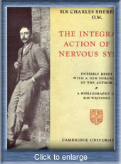
In 1906 he published his well-known book: The Integrative Action
of the Nervous System, being his Silliman Lectures held at Yale University
the previous year, and in 1913 he was invited to become Waynfleet
Professor of Physiology at Oxford, a post for which he had unsuccessfully
applied in 1895, and here he remained until his retirement in 1936. Here
he wrote, and published in 1919, his classic book entitled Mammalian Physiology:
a Course of Practical Exercises, and here he regularly taught the students
for whom this book was written.
The predominant notes of his character as a man were his humility and friendliness and the generosity with which he gave to others his advice and valuable time. An interesting feature of him is that he published, in 1925, a book of verse entitled The Assaying of Brabantius and other Verse, which caused one reviewer to hope that «Miss Sherrington» would publish more verse. He was also sensitive to the music of prose, and this and the poet in him, but also the biologist and philosopher, were evident in his Rede Lecture at Cambridge in 1933 on The Brain and its Mechanism, in which he denied our scientific right to join mental with physiological experience.
The philosopher in him ultimately found expression in his great book, Man on his Nature, which was the published title of the Gifford Lectures for 1937-1938, which Sherrington gave. As is well known, this book, published in 1940, centres round the life and views of the 16th century French physician Jean Fernel and round Sherrington's own views. In 1946 Sherrington published another volume entitled The Endeavour of Jean Fernel.
Nobel prize for medicine in 1932 with Lord Edgar Douglas Adrian .

Drawing of overlapping neuronal fields by Sherrington
Adrian's first research work was done with Keith Lucas, who was working on the impulses transmitted by motor nerves; he showed that, when a muscle fibre contracts, the passage of the nerve impulse that causes the contractions leaves the motor nerve in a state of diminished excitability. Keith Lucas was, at the time of the First World War, thinking of improving the study of the electrical currents in nerves by amplifying them by means of valves, a method which Adrian was later to employ. In order to obtain a more sensitive detection of nerve impulses, he used the cathode ray tube, the capillary electrometer and amplification of the electrical impulses by means of thermionic valves, and was thus able to amplify them 5,000 times. He developed fine needle electrodes, using the tip of a needle to record from specific muscles. With this apparatus he was able to record the electrical discharges in single nerve fibres which were produced by tension on the muscle, pressure on it, touch, the movement of a hair and pricking with a needle. By 1928 he was able to publish his conclusion that a stimulus of constant intensity applied to the skin, immediately excites the end organ, but that this excitation progressively decreases for as long as the stimulation continues. At the same time sensory impulses of constant intensity pass along the nerve from the end organ.
For his work about the functions of neurones Adrian was awarded, jointly with Sir Charles Sherrington, the Nobel Prize for 1932.
The Basis of Sensation (1927)
The Mechanism of Nervous Action (1932)
The Physical Basis of Perception (1947).
Adrian ED, Bronk DW (1928) The discharge of impulses in motor nerve
fibers. Part II: the frequency of discharge in reflex and voluntary contractions.
J Physiol. 66:81-101.
The shoulder-piece is supported laterally on springs, of suitable
strength, which act as shock-absorbers, and bear the
weight of the body, imparting to it a sort of oscillation, which
accelerates the movement of propulsion and lessens the
axiallary pressure.... The wounded soldier must be led, by successive
stages, to rely less and less on the double support
of the armpits, and to employ the muscles of the limbs, without
fatiguing them. That he does not do this I am able to
assure myself by measuring the respiratory energy.
The crutch was a kind of self-therapy; its tremendous power lay in its capacity to make the maimed soldier an active participant in his own rehabilitation - a single unit of the worker and the machine, a kind of early cyborg whose biological component was conditioned by the mechanical part.
Amar argued that the same science should apply to the treatment of wounded
soldiers and ordinary workers, because both
endeavors sought "the geometrical and harmonious forms of the contractions
of the muscles."[38] He sought to bind together
the soldier and the worker on the basis of their common physiology:
If, in order to execute any physical action whatever...we eliminate the useless movements, and regulate the succession of the useful, we shall effect a great saving both of time and fatigue.... Selection and order are, in truth, the characteristics of the new method, which will presently work an economic revolution to which no other can be compared. It is not purely mechanical; it does not turn a man into a soulless body, a blind and tireless force; it embraces all the data of physiology and psychology, of which it alone is able to display the parallelism and the unfailing harmony. It would seem to have taken for its guide this saying of Montaigne's: "It is not a body, it is not a soul that we are forming: it is a man; we must not make two of him
Amar J (1920) The Human Motor
Amar J (1920) The Physiology of Industrial Organization and the Re-Employment
of the Disabled
 Russell
Plato Schwartz, M.D. (1892-1965), earned his medical degree from Indiana
University (1920). Appointed as a teacher in surgery, R. Plato Schwartz
was in time promoted to head of orthopedic surgery at the University of
Rochester School of Medicine & Dentistry from 1926 until his retirement
in 1957. He was the first clinician to develop instrumented gait analysis
for clinical assessment.
Russell
Plato Schwartz, M.D. (1892-1965), earned his medical degree from Indiana
University (1920). Appointed as a teacher in surgery, R. Plato Schwartz
was in time promoted to head of orthopedic surgery at the University of
Rochester School of Medicine & Dentistry from 1926 until his retirement
in 1957. He was the first clinician to develop instrumented gait analysis
for clinical assessment.
The Myodynamics Laboratory (or 'Gait Laboratory') was established within the Department of Surgery in 1926, soon after his appointment as Assistant Professor of Orthopedic Surgery. Dr. Schwartz's intention was to devise mechanisms for the accurate recording of human locomotion in order to establish norms for both normal and abnormal gait. The Laboratory's first efforts resulted in a pendulum basograph in 1926, which by the early thirties had been replaced by a pneumographic gait recorder (on the principle of Marey's). On June 12, 1933, an electrobasograph had been constructed to measure "the walking gait of individuals, to distinguish between actual and spurious limps in damage claims for injuries." It was exhibited to the American Medical Association convention in Milwaukee, Wisc.
In the 1930s the Myodynamics Laboratory began its association with the
Armstrong Shoe Company of Rochester, N.Y., who were interested in the application
of
the Laboratory's research on foot function to shoe design. This began
the first of a long series of contacts with American and European last
and shoe manufacturers,
and quickly became the Laboratory's principle source of income.
An overzealous U. of R. publicity man circulated a story, which Time magazine (Sep. 27, 1943) seized upon, that Schwartz contemplated applying his orthopedic knowledge to making race horses trot faster. A foundation which had appropriated funds for scientific investigations at the U. of R., and the American Medical Association as well, objected strenuously to this form of research and the attendant advertising; the publicity agent was ordered by his superiors to cease and desist. Schwartz also worked as a consultant to the national government on military footwear, and at about mid-point in his career he organized the first cerebral palsy center in New York State--the Edith Hartwell Clinic at LeRoy, New York--and was invited to lecture in Europe on this phase of health care. In the late 1940s and 1950s, the Laboratory extended its prior electromyographic researches into the effects of poliomyelitis and cerebral palsy on muscle function.
In 1931 Arthur L. Heath, B.S. joined the Myodynamics Laboratory as a
Research Associate. His association with the Laboratory lasted through
its closing in 1969,
by which time he was Associate Director. From the mid-1930s, research
in the Myodynamics Laboratory focused increasingly on the development of
"functional" principles of last and shoe design, with the continuing perfection
of gait recording instruments and the development of such mechanisms as
the mirrorscope, the last balograph and the last contourgraph. The Laboratory
maintained its interest in the pure mechanics of human locomotion, however,
and the application of these studies to the diagnosis and correction of
gait abnormalities, whether caused by injury or congenital condition.
In conjunction with Dr. Schwartz's separate researches into poliomyelitis
and cerebral palsy The Laboratory was also instrumental in the founding
of the
Edith Hartwell Clinic in Leroy, N.Y., a faciltity for the training
of children affected with cerebral palsy.
After Dr. Schwartz's retirement in June 1957, the Myodynamics Laboratory
continued under the direction of Arthur Heath. Despite Mr. Heath's attempts
in the
mid-1960s to organize a Foot & Footwear Research Institute on the
foundations of the Myodynamics Laboratory, the program appears to have
been gradually
phased out and is last mentioned in the 1968/69 issue of the Official
Bulletin of the School of Medicine & Dentistry.
Various medical associations bestowed medals on him for his diversified achievements
Schwartz, R.P., Vaeth, W. "A method for making graphic records of normal
& pathologic gaits," JAMA, Jan. 1928.
Schwartz, R.P., & Tuttle, H.B. "Advantages of using 16mm Cinekodak
super-sensitive panchromatic film in making medical motion pictures," J.
Soc. Motion Pict. Engineers, v. 18, 1932; & NYS j. med., Oct. 1932.
Schwartz, R.P. "The pneumographic method of recording gait," J. bone
& joint surg., Oct. 1932.
Schwartz, R.P., Heath, A.L., & Wright, J.N. "Electrobasographic
method of recording gait," Arch. surg., Nov. 1933.
Schwartz, R.P., Heath, A.L., & Misiek, W. "The influence of shoe
on gait," J. bone & joint surg., Apr 1935.
Schwartz, R.P., & Heath, A.L. "The feet in relation to the mechanics
of human locomotion," Physiother. rev., Mar-Apr 1936.
Schwartz, R.P., Trautmann, O., & Heath, A.L. "Gait and muscle function
recorded by the electrobasograph," J. bone & joint surg., Apr 1936.
Schwartz, R.P., & Heath, A.L. "Some factors which influence the
balance of the foot in walking," J. bone & joint surg., Apr 1937.
Schwartz, R.P., & Heath, A.L. "The definition of human locomotion
on the basis of measurement," J. bone & joint surg., Jan 1947.
Schwartz, R.P., Heath, A.L., & Hudson, F.W. "Instrumentation in
relation to electro- myography [Pt. 2]," Arch. phys. med., JuneSchwartz,
R.P., & Heath, A.L. "The oscillo- graph recording and quantitative
definition of functional disabilities of human loco- motion," Arch. phys.
med., Sep 1949.
Schwartz, R.P., Heath, A.L., et al. "A quan- titative analysis of recorded
variables in the walking pattern of 'normal' adults," J. bone & joint
surg., Mar 1964.
 The Russian physiologist
Bernstein challenged the view held in McGraw's and Gesell's time of a hierarchical
system within the body whereby commands for movement were issued by the
brain. He posited that performance of any kind of movement results
from an infinite variety of possible combinations, or degrees of freedom,
of neuromuscular and skeletal elements. The system should, therefore,
be considered as self-organizing, with body elements coordinated, or assembled,
in response to specific tasks. Motor development was dependent not
on brain maturation, but adaptations to constraints of the body (changes
in the growing infant's body mass and proportions) and to exogenous conditions
(gravity, surface, specific tasks to be performed). biomechanics.
Bernstein coined the term biomechanics, describing the application of mechanical
principles and methods to biological systems. His book Coordination
and Regulation of Movements was not published in the West until 1967.
Many of the key issues in modern day movement coordination were formulated
in this text, including the degree of freedom problem, motor equivalence,
and non-univocality of motor commands and peripheral effects.
The Russian physiologist
Bernstein challenged the view held in McGraw's and Gesell's time of a hierarchical
system within the body whereby commands for movement were issued by the
brain. He posited that performance of any kind of movement results
from an infinite variety of possible combinations, or degrees of freedom,
of neuromuscular and skeletal elements. The system should, therefore,
be considered as self-organizing, with body elements coordinated, or assembled,
in response to specific tasks. Motor development was dependent not
on brain maturation, but adaptations to constraints of the body (changes
in the growing infant's body mass and proportions) and to exogenous conditions
(gravity, surface, specific tasks to be performed). biomechanics.
Bernstein coined the term biomechanics, describing the application of mechanical
principles and methods to biological systems. His book Coordination
and Regulation of Movements was not published in the West until 1967.
Many of the key issues in modern day movement coordination were formulated
in this text, including the degree of freedom problem, motor equivalence,
and non-univocality of motor commands and peripheral effects.
Berstein compared the running technique of the famous Zhamenski brothers and the then world record holder, Frenchman, Jules Ladoumegue. The French champion seemed to be landing his feet near the imaginary line, going through the General Center of Mass perpendicular to the track. The Soviet athletes, on the other hand were throwing their feet far forward and later were "crashing down" into them, and losing their speed. On the basis of exactly these long-term research studies, Nicholas Romanov developed his running method, which he uses today to train the British Olympians.
Bernstein, and his coworkers at the Russian All-Union Institute of Experimental Medicine in Moscow, conducted an extensive investigation of body segment parameters of living subjects. A total of 152 subjects of both sexes, ranging in age from 10 to 75 years were examined and the mass and center of mass of all limb, excluding the center of mass of hands and feet, were estimated. The estimations of the mass of body segments were performed using a modified balance plate. The balance plate technique used by Borelli had been modified and improved several times over the years and a simplified sketch of the version used by Bernstein is shown in the figure.
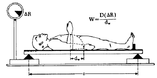
The system can be used to establish a relation between the mass and
the displacement of the center of mass of a body segment. The relation
is given by W =D(DR) dw , where W is the mass of the body segment, D is
the distance between the two supporting knife edges, dw is the displacement
of the center of mass of the body segment and DR is the change in pressure
exerted on the scale due to this displacement (see Figure 1). The problem
that remains is that neither the center of mass nor the mass of the segment
easily and accurately can be estimated by other methods. Bernstein concluded,
however, by examining frozen cadaver segments, that the center of mass
of a segment, for most practical purposes, coincides with the center of
volume. Assuming this coincidence and since the volume and center of volume
of a segment can be estimated in vivo, then the center of mass and subsequently
the mass of the body segments of living subjects can be estimated. Bernstein
concluded, that the individual variations was so great that either complex
measuring techniques, as the ones described above, should be used on every
individual subject that is dealt with, or anthropometric and structural
correspondences (correlations), which allow estimations to be performed
based on general habits and anthropometric data, should be established.
Bernstein NA (1967) The Co-ordination and Regulation of Movements, Pergamon
Press, Oxford
Bernstein NA (1927) Untersuchungen der Biodynamik des Ganges und des
Laufes. Moscow
Bernstein NA (1935) Untersuchungen der Biodynamik der Lokomotion. Moscow
Benstein NA (1988) Bewegungsphysiologie, Leipzig
Benstein NA (1991) Über die Gewandtheit und ihre Entwicklung,
Moskau
Benstein NA (1996) Die Entwicklung der Bewegungsfertigkeiten, Leipzig
 Vulpius-Freiluftsanatorium,
founded 1912
Vulpius-Freiluftsanatorium,
founded 1912
In 1912, with Adolf Stoffel, the Heidelberg professor Dr. Oskar Vulpius established a 120 bed orthopaedic hospital, Sanatorium Solbad bath Rappenau, located on the edge of the forest in the spa town of Rappenauer. Gastrocnemius lengthening for isolated contracture of the gastrocnemius was pioneered by Vulpius and Stoeffel.
Vulpius, O & Stoffe A (1913) Orthopadische operationslehre. Stuttgart
: Ferdinand Enke.


Elftman's mechanical force platform, capable of recording the shear components of the ground reaction, as well as the load, via the displacements of five levers, which were filmed by high-speed cine camera, which simultaneously recorded the kinematics.
Elftman H (1934) A cinematic study of the distribution of pressure in
the human foot. Anat. Rec. 59: 481-487.
Elftman
H (1938) The measurement of the external force in walking. Science 88:152-153.
Elftman H (1939) Forces and Energy Changes in the Leg During Walking.
Am. J Physiol. 25:339-356.
Dudley Morton lent his name to Morton's syndrome (not the neuroma, incidentally, which is due to Thomas G. Morton). This is characterized by a short first metatarsal bone causing excessive weight to be borne by the second metatarsal head. It is usually a hereditary condition and will result in callus formation under the second and third metatarsals. Pain and tenderness are usually felt at the base of the first two metatarsal bones and at the head of the second.
Morton, and an orthopaedic surgeon, Paul Lapidus, independently published similar theories that implicated excessive mobility of the first metatarsal in forefoot dysfunction. Morton first studied in detail the evolution of the foot leading to bipedal gait and then examined the feet of many of his students in an attempt to correlate his tentative assumptions with the presence of or lack of symptoms. Morton's analysis of forefoot dysfunction provides the foundation of modern surgical procedures that restore normal forefoot balance and function to asymptotic feet.
Evolution of the Human Foot (1922) Am J Anthropology 5(4): 305-336 &
7(1): 1-52.
Evolution of the longitudinal arch of the human foot (1924) J Bone
& Joint Surgery 6: 56-90.
Structural factors in static disorders of the human foot (1930) Am
J Surgery 9: 315-326.
Human locomotion and body form. A study of gravity and man (1952) Williams
and Wilkins, Baltimore.

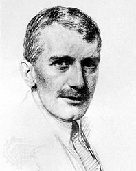
Born in Bristol, educated at Blundell's School, Tiverton, whence he obtained scholarships to Trinity College, Cambridge. Here he studied mathematics and took the Mathematical Tripos, being Third Wrangler (1907). After graduating, he was urged to take up physiology by his teacher, Dr. (later Sir) Walter Morley Fletcher.
Hill started his research work in 1909. It was due to J. N. Langley, Head of the Department of Physiology at that time that Hill took up the study on the nature of muscular contraction. Langley drew his attention to the important (later to become classic) work carried out by Fletcher and Hopkins on the problem of lactic acid in muscle, particularly in relation to the effect of oxygen upon its removal in recovery. During his initial studies Hill made use of the Blix' apparatus, obtaining his first knowledge of the subject from papers of this Swedish physiologist. This led him to study the dependence of heat production on the length of muscle fibre (a relation later developed by Starling in his investigation of the mechanism of the heart beat).
After having obtained a Fellowship at Trinity in 1910, Hill spent the
winter of 1910-1911 in Germany, working among others with Bürker (who
taught him much about the technique of myothermic observations) and Paschen
(who introduced the galvanometer to him, which he since used for his investigations).
From 1911-1914, until the outbreak of World War I, he continued his work
on the physiology of muscular contraction at Cambridge. During this for
him important period, however, he also took up other studies: on the nervous
impulse (with Keith Lucas), on haemoglobin (with Barcroft), and on calorimetry
of animals (partly with T. B. Wood), having also as colleagues Gaskell,
Anderson, W. B. Hardy, Mines, Adrian, Hartridge, and others.
In 1919 he took up again his study of the physiology of muscle, and came into close contact with Meyerhof of Kiel who, approaching the problem from a different angle, has arrived at results closely analogous to his study. They have cooperated continuously ever since, by personal contact and through correspondence. In 1919 Hill's friend W. Hartree, mathematician and engineer, joined in the myothermic investigations - a cooperation which had rewarding results.
In 1920 Hill was appointed Brackenburg Professor of Physiology at Manchester University; there he continued the work on muscular activity and began to apply the results obtained on isolated muscles to the case of muscular exercise in man. From 1923 to 1925 he became Jodrell Professor of Physiology at University College, London, succeeding E. H. Starling. In 1926 he was appointed the Royal Society's Foulerton Research Professor and was in charge of the Biophysics Laboratory at University College until 1952.
With Meyerhof, he received the 1922 Nobel prize for ""for his discovery relating to the production of heat in the muscle"
Muscular Activity (1926)
Muscular Movement in Man (1927)
Living Machinery (1927)
Hill, A.V. (1938). The heat of shortening and the dynamic constants
of muscle. Proc. R. Soc, London B, 126, 136-195.
Hill, A.V.: The series elastic component of muscle, Proc. R. Soc. Lond.
(Biol.) 137: 273-280, 1950.
Hill, A.V.: The mechanics of active muscles, Proc. R. Soc. Lond. (Biol.)
141:104-117, 1953.
Fenn, W.O., & Marsh, B.S. (1935). Muscular force at different speeds of shortening. Journal of physiology, 85(3), 277-297
Responsible for some of the greatest explanations of how muscles physically contract. In 1953, he revealed his sliding filament theory to explain muscle contraction, and following this he postulated his famous Cross-bridge Theory to further explain the mechanism of muscle action. His application of physics to muscle physiology was also responsible for many other accomplishments such as the development of X-ray diffraction and electron microscopy.
Huxley, A.F. and Niedergerke, R. (1954). Structural changes in muscle
during contraction. Interference
microscopy of living muscle fibres. Nature, 173, 971-973.
Huxley, A.F. (1957). Muscle structure and theories of contraction.
Progress in Biophysics and Biophysical Chemistry 7. 255-318.
Hodgkin, A. L. and Huxley, A. F. (1952) "A Quantitative Description
of Membrane Current and its Application to Conduction and Excitation in
Nerve" Journal of Physiology 117: 500-544
In 1953, in collaboration with Jean Hanson, Huxley proposed the sliding-filament theory of muscle contraction. This was based on his earlier study of myofibrils, the contractile apparatus of muscle, with the electron microscope. He found that myofibrils are made of two kinds of filament, one type about twice the width of the other. Each filament is aligned with other filaments of the same kind to form a band across the myofibril, and the bands of thick and thin filaments overlap for part of their length. The bands are also linked by an elaborate system of crossbridges. When the muscle changes length the two sets of filaments slide past each other. Further, the two sorts of filaments can be identified with the two chief proteins of muscle, myosin in the thick filament and actin in the thin. This made possible an elegant solution to how muscles contract at the molecular level.
In the areas where both kinds of protein are in contact, Huxley suggested that one, most probably myosin, serves as an enzyme, splitting a phosphate from ATP and so releasing the energy required for contraction. He concluded that the evidence of the combination of actin and myosin is seen in the bridges between the two kinds of filaments.
The theory has since been much enlarged and taken to deeper levels of molecular understanding. Despite this, the basic insight of Huxley and Hanson has remained intact.
Huxley, H. E. 1969. The mechanism of muscle contraction. Science 164:
1356-1365.