Gait Analysis Models in Common Use
Mathematics is so different when applied to people. One plus one can
add up to so many different things
Werner Heisenberg
|
Parameter
|
Helen Hayes
Vicon VCM (Roy Davis)3
|
GaitLab
Peak (Kit Vaughan)
|
Cleveland Clinic
MAC Orthotrak (Kadaba)
|
QualiSys
|
Ariel
|
Elite
|
| Type |
Anthropometric (using very limited set of measurements) |
Anthropometric (using detailed set of measurements) |
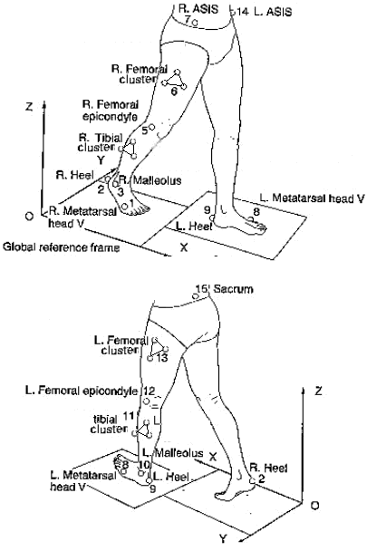
Cluster |
|
|
|
| Marker Set |
 |
 |
 |
|
|
|
| Anthropometry |
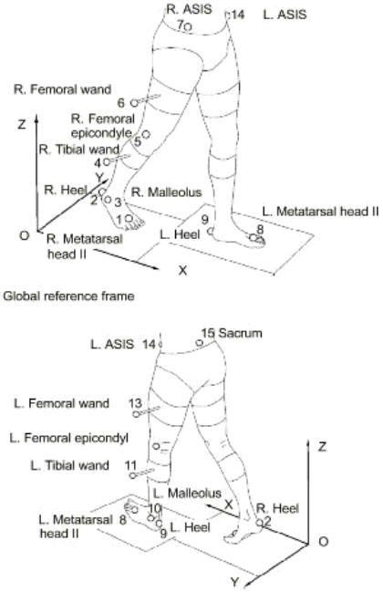
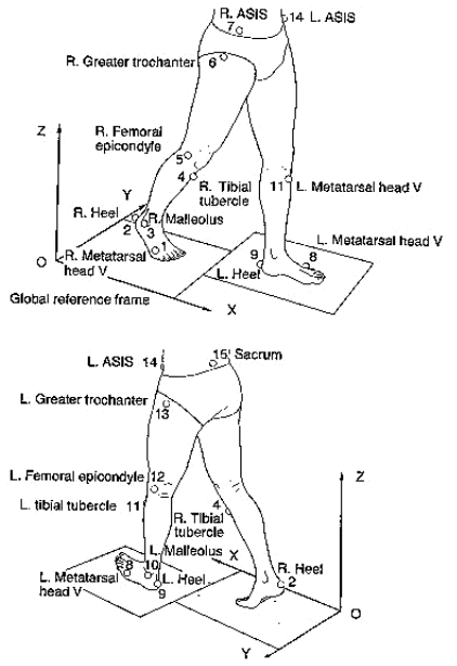
Leg Length (ASIS-Med Mall)
Knee Width
Ankle (inter-malleolar) Width |
Inter-ASIS distance
Asis Breadth
Thigh Length
Midthigh Circumference
Calf Length
Calf Circumference
Knee Diameter
Foot Length
Malleolus Height
Malleolus Width
Foot Breadth |
|
|
|
|
Calibration
(Static Trial) |
User-defined torsion values for femur and tibia
Heel (calcaneal) markersduring static trial to determine ankle
offset angles
Optional Knee Alignment Device (KAD) to assist in femoral wand placement |
|
User defines thigh orientation by indicating knee axis |
|
|
|
| Hip Joint Center |
C = LegLength*0.115-15.3
InterASISDist=DIST(LASI,RASI)
aa = InterASISDist/2
mm = MarkerDiameter/2
ATD = AsisTrocanterDistance
COSBETA = 0.951
SINBETA = 0.309
COSTHETA = 0.880
SINTHETA = 0.476
HJC = {C*COSTHETASINBETA - (ATD + mm) * COSBETA,
-C*SINTHETA + aa,
-C*COSTHETACOSBETA - (ATD
+ mm) * SINBETA}*Pelvis
Davis RB et al. A gait analysis data collection and reduction technique.
Human movement science 1991(10): 575-587 |
Sacral Marker
+0.598(ASIS breadth)upelvis
+/-0.344(ASIS breadth)vpelvis
-0.290(ASIS breadth)wpelvis |
|
|
|
|
| Knee Joint Center |
KJC=CHORD(KneeOS,KNE,HJC,THI)
KneeOS = (MarkerDiameter+KneeWidth)/2 |
femoral epicondyle marker+0.5(Knee diameter) |
indicated by user with wand |
|
|
|
| Ankle Joint Center |
AJC=CHORD(LAnkleOS,ANK,KJC,TIB) |
Lateral malleolus marker
+0.016(Foot length)ufoot
+0.392(Malleolus height)vfoot
+0.478(Malleolus width)wfoot |
|
|
|
|
| Toe Point |
|
Lateral malleolus marker
+0.742(Foot length)ufoot
+1.074(Malleolus height)vfoot
-0.187(Malleolus width)wfoot |
|
|
|
|
| Body Center of Mass |
If $age>14 then <Dempster anthropometry>
CoM=(0.078*HeadCoM)
+(0.027*LHumerusCoM)
+(0.027*RHumerusCoM)
+(0.023*LForearmCoM)
+(0.023*RForearmCoM)
+(0.503*TrunkCoM)
+(0.099*LFemurCoM)
b+(0.099*RFemurCoM)
+(0.046*LTibiaCoM)
+(0.046*RTibiaCoM)
+(0.014*LFootCoM)
+(0.014*RFootCoM)
else <Jensen anthropometry>
CoM=((0.238-0.0114*age)*HeadCoM)
+((0.00084*age+0.022)*LHumerusCoM)
+((0.00084*age+0.022)*RHumerusCoM)
+((0.00015*age+0.012)*LForearmCoM)
+((0.00015*age+0.012)*RForearmCoM)
+((-0.0006*age+0.4246)*TrunkCoM)
+((0.00364*age+0.06634)*LFemurCoM)
+((0.00364*age+0.06634)*RFemurCoM)
+((0.00122*age+0.03809)*LTibiaCoM)
+((0.00122*age+0.03809)*RTibiaCoM)
+((0.00015*age+0.0187)*LFootCoM)
+((0.00015*age+0.0187)*RFootCoM)
Endif |
|
|
|
|
|
Notes
1. The operators * and / have a special meanings in VCM
point operations. They are used to transform a point between its global
version in the VICON calibrated space and its "local" version in a segment,
and vice versa;
e.g. pointI = pointI*segmentP converts local pointI in segmentP to global
2. u, v, w are the unit vectors of a segment in the
x, y, z directions
3. The VCM model
The marker set is ASIS, Sacrum (S2, between the skin
dimples), thigh and shank wands (more of this later),
lateral femoral condyle (this can be tricky - more
later), lateral malleolus and second metatarsal head
(just proximal to the MTPJ).
The thigh wand and knee marker are the most tricky
elements. Together with the virtual hip joint (derived
by anthropometry) they define both the knee joint
center and frontal plane of the thigh.
I used to place the knee marker so that its forward
edge (1 inch diameter) bisects the dorsal and ventral
aspects of the joint - a common error is to place it
too anterior.
The thigh wand (a single marker on a 4 inch stick)
needs to be placed in a line with this marker and the
hip joint, with the subject standing at the side of
the operator. Of course, this is easy to say but
difficult to do in practice because:
a. You don't actually know where the hip joint is!
b. You're so close to the subject that you can't
easily determine when the three markers are co-linear.
We talked about this at length on CGA - see:
/faq/cleveland.html
The method I used was to place
a mirror on the wall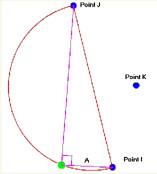
several yards away, and turn the subject so that the
leg being markered was seen in the mirror. I then
placed a finger where I thought the hip jont was
(greater trochanter for normals - you have to use you
intuition for patients!) and then adjust the wand
until it formed a straight line with the finger and
knee joint marker. I git this from Jeremy Lindskill in
Dundee, although it's interesting that all his data
has a large knee flexion offset because he's always
placed the marker too anterior - by the time he
realised he had collected too much data, and has
decided to just carry on collecting erroneous data for
compatibility reasons!
I know this sounds ridiculous - it's the major problem
with VCM. If you get the thigh wand wrong, you will
get big varus/valgus artifacts, and I have seen this
on most people using Vicon (a lot of people don't even
know about it - one doctor on CGA actually launched
into a detailed interpretation of a huge varus
artifact!). As the software improved what I was able
to do was take a quick trial and look for artifacts -
then adjust the markers accordingly before collecting
real data. This might be one possible advantage of
real time.
In the software Julian invented what he calls a
"chord" function, which simply draws an arc based on
the calculated hip joint center and passing through
the knee marker and thigh wand. It then draws a chord
through this arc at the knee marker, which is the
computed knee joint axis. Halfway along this is the
center.
CHORD(numberA,pointI, point J, pointK)
Point at distance A from I in plane IJK forming a right angle between
I and J on the opposite side of IJ from K
Note that this was all done at the time to save on
markers - the early systems had trouble tracking more
than a dozen or so (VCM has 13 markers).
BTW, there are also two more markers added on each
calcaneus at the level of the toe markers. These are
only used for the static trial to calculate the ankle
offset angle, and can be removed for the actual data
collection.
References
Davis, RB III, Ounpuu, S, Tyburski, D, and Gage, JR (1991). A gait data
collection and reduction technique. Human Movement Sciences 10, 575-587.
Kadaba M P, et al. (1989). Repeatability of Kinematic, Kinetic and Electromyographic
Data in Normal Adult Gait. Journal of Orthopaedic Research, 7:6, 849-860.
Tabakin, D. & Vaughan, C.L. 2000. A comparison of 3D gait models
based on the Helen Hayes marker set, Proceedings of the Sixth International
Symposium on
the 3D Analysis of Human Movement, Cape Town, South Africa, 98-101.
Vaughan CL, Davis BL, O'Connor J (1992) Dynamics of Human Gait. Human
Kinetics Press, Champaign Illinois - see also GaitCD
and GaitLab
(2nd edition of Gait Analysis Laboratory)