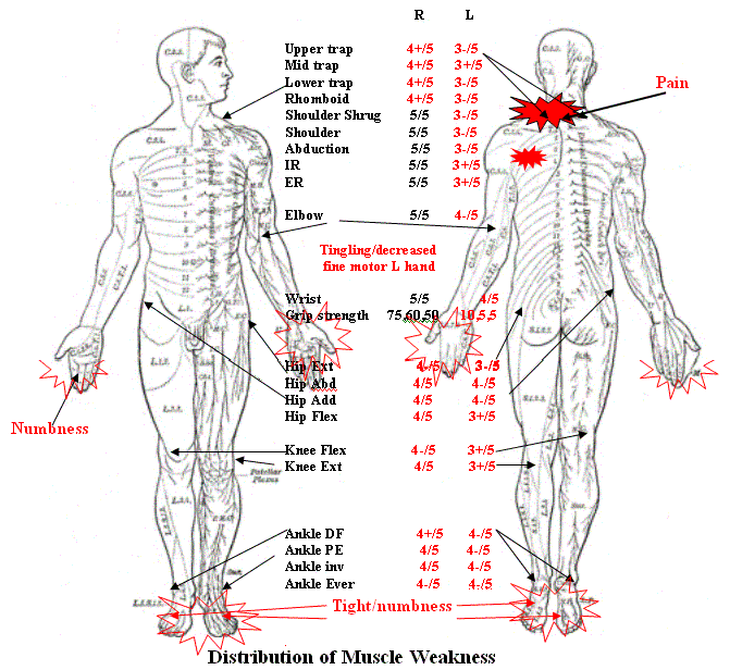
• Pain mid-cervical to top of left shoulder
• Left shoulder blade pain
• Numbness top of left shoulder
• Tingling left hand, mostly 1st three fingers with decreased sensation
• Occasional color change in left hand - mottled appearance
• Difficulty spreading fingers apart on left with impaired fine motor control, occasionally dropping items
• Weakness left arm
• Unable to raise arm higher than level of shoulder - as if shoulder was paralyzed
• Unable to lift half gallon of milk or heavier object
• Muscle twitching noted mostly left biceps, triceps and left upper thigh area initially - now mostly left side, with occasional twitching in right arm
• Numbness in right hand periodically - sometimes whole hand including 5th finger (occurs if lying on either right or left side), or just right thumb
• Numbness left toes March 2003Notes
• Slower walking July 2003
• Numbness right toes August 2003
• Limping started September 2003
• Weakness left leg more apparent to patient October 2003
• Bilateral toe sensation like “shoes too tight”
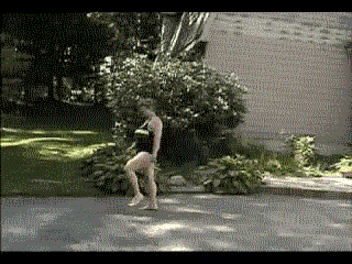
- Walking increasingly difficult ever since with increased difficulty going up and down stairs
- Gait is abnormal, high stepping, decreased endurance, balance on R good, on L fair.
Neck pain greatly reduced with in supine position, but hand tingling and shoulder blade pain worse Arm pain present when head thrust to left or head tilted to left and axially compressed Bumpy car rides increase pain and tingling Position of head to right side while lying flat corrects shoulder problem (not possible if up all day). Initially if my head was flat and slightly hyperextended I could raise my arm completely over head and lift heavy object-unable to duplicate this for over a year. Bilateral foot tightness sensation not present first thing in morning after lying supine all night - but commence immediately on rising
November 14, 2002 Cervical MRI: asymmetrical bulging on left at C5-6 but no compression seen, most likely post-op changes
December 2, 2002 EMG done by Neurology: C7 Radiculopathy acute and chronic.
December 18, 2002 CT Myelogram: some C7 anterior sleeve filling defect, probably post-op in nature.
January 15, 2003 Cervical spine x-rays: no instability
January 23, 2003 EMG (results [verbal] identical to EMG performed 12/02/02-active C7 radiculopathy
February 14, 2003 cranial MRI: normal
June 20, 2003 Stand up MRI: Bulging at 2-3, 3-4 impinging on thecal sac, posterior central herniation impinging on cord, minimal narrowing, ventral and dorsal marginal osteophytes, diffuse bulging at 5-6, 6-7 impinging on ventral thecal sac and minimal narrowing of 5-6, 6-7 neural foramina bilaterally, straightening and desiccation of all disks. T4 and 5 vertebral body lesions probably representing atypical hemangiomas
July 9, 2003: “unofficial report” from neuroradiologist: Straight cervical spine, hypermobile intersegmental instability C4-5. posterior central herniation c4-5 larger with extension, herniation at C5-6 to left neural foramina, herniation C6-7 to left neural foramina, larger with extension. C7 nerve root enhancement ? benign neural inflammation ? Herniations smaller with flexion.
August 28, 2003: second positional Cervical MRI
October 8, 2003 CT and cervical spine studies
Cervical spine results: degenerative disease C5-6-7. Small spurs left
side increased encroachment during flexion compared to extension, limited
range of motion, no spondylolisthesis
CT results-small posterior and central disc protrusion C4-5, degenerative
disease C5-6/C6-7 with diffuse annular disc bulges and osteophyte formastion,
mild encroachment upon neural foramina due to associated uncovertebral
joint osteophyte formation
October 30, 2003: MRI in very specific postitions: head tilting towards
left side-very painful in left scapula area, down left arm with increased
tingling. Gait feeling unsteady and weak. Pain and arm weakness unchanged.
Unofficial report: degenerative osteophyte formation C5-6 C6-7 extending
into neural foramina.
December 16,2003 EMG: normal
December 18, 2003 SSERs: normal
February 23, 2004: ANA positive, all other lab work negative. Lupus and other auto immune, rheumatologic diseases excluded.
May 12, 2004 MRI report: thoracic spine - within normal limits, normal signal intensity within the thoracic spinal cord, no sign of injury or demyelination. Lumbar - mild dengenerative broad annular disc bulges from L3-4 through L5-S1 without central canal or neural foraminal narrowing, no focal disc herniation. Mild hypertrophic degenerative facet spondylosis.
May 20, 2004 B&Ws: EMG left arm and leg. Impression - Left H reflex absent (Leg Study - three attempts to elicit response) while right H reflex present and normal. “Active, ongoing denervation, complex repetitive discharges and chronic reinervation in the form of long units in the pronator teres. May represent a chronic C7 radiculopathy. Absent H reflex is non-specific finding of uncertain clinical significance
June 4, 2004 Cranial MRI: normal
July 16, 2004 Complete Evoked Potentials, including auditory, visual and bilateral motor of all extremities: all normal.
September 7, 2004 Laboratory tests pending, including Ceruloplasmin, C-Reactive Protein, Creatine Kinase, Lyme Ab, Paraneoplastic Ab, Purkinje Cell Ab, Protein Electrophoresis, RPR, Vitamin B12, Gliadin Ab, Vitamin E.
Movies
AVI format
|
|
front | back |
| Sagittal | right | left |
![]() Points for discussion
Points for discussion
Email your comments to [n/a]